Wednesday, April 30, 2008

Suture Allergies Revisited

Updated 3/2017-- photos and all links removed as many are no longer active and it was easier than checking each one.

Over the last few months I have received several e-mails from patients with suture reactions, possibly allergic reactions. I say possibly because not being their treating physician I can't verify them, but by the descriptions they seem real. I reviewed this topic last year, but felt the need to revisit it. Hope you don't mind me sharing with you.
Allergic reactions to suture materials are rare and have been specifically associated with chromic gut. However, Johnson and Johnson mention known triclosan allergy as a contraindication for use of certain sutures (see below). Contact allergy to triclosan is uncommon.
Surgical gut suture (Plain and Chromic) is contraindicated in patients with known sensitivities or allergies to collagen or chromium, as gut is a collagen based material, and chromic gut is treated with chromic salt solutions.
MONOCRYL Plus Antibacterial suture should not be used in patients with known allergic reactions to Irgacare MP(triclosan).
PDS Plus Antibacterial suture should not be used in patients with known allergic reactions to Irgacare MP (triclosan).
VICRYL*suture should not be used in patients with known allergic reactions to Irgacare MP (triclosan).
Surgical Stainless Steel Suture may elicit an allergic response in patients with known sensitivities to 316L stainless steel, or constituent metals such as chromium and nickel. Skin staples are surgical steel so should be used with the same precautions.
Dermabond -- Tissue glues should not be used in patients with a known hypersensitivity to cyanoacrylate or formaldehyde.

SO WHAT IS LEFT TO USE
So what is left to use in a patient who may have or has a proven allergy to suture or closure material?
Silk, Dexon, Nylon(monofilament or braided), Prolene, INSORB (absorbable staples), and any of the above listed (in the allergy section) to which the patient in question doesn't react negatively.
The choice of a particular suture material will have to based further on the wound, tissue characteristics, and anatomic location. Understanding the various characteristics of available suture materials will be even more important to make an educated selection.
The amount of suture placed in a wound, particularly with respect to the knot volume, affects inflammation. The suture size contributes more to knot volume than the number of throws. The volume of square knots is less than that of sliding knots, and knots of monofilament sutures are smaller than those of multifilament sutures.
REFERENCES
  • Allergic Suture Material Contact Dermatitis Induced by Ethylene Oxide: G. Dagregorio, G. Guillet; Allergy Net Article
  • Current Issues in the Prevention and Management of Surgical Site Infection - Part 2; MedScape Article
  • MECHANICS OF BIOMATERIALS: SUTURES AFTER THE SURGERY; Raúl De Persia, Alberto Guzmán, Lisandra Rivera and Jessika Vazquez
  • Materials for Wound Closure by Margaret Terhune, MD; eMedicine Article
  • Product Allergy Watch: Triclosan; MedScape Article by Lauren Campbell; Matthew J. Zirwas

Tuesday, April 29, 2008

Grand Rounds--Are You Ready to Rumble?

 Updated 3/2017-- photos and all links removed as many are no longer active and it was easier than checking each one.

This weeks edition : Grand Rounds: Smack Down Week over at Doc Gurley's.
“Are you ready to ruum-ble? This week here at Doc Gurley is the Grand Rounds Smack Down edition, where the best contenders of the health care blogosphere wrestle down and dirty with tough, scary topics. Just like being in the ring, there’s no orderly progression of characters here – this ain’t ballet, folks. At any moment, something huge and impressive could come flying out of left field and hit you (ka-pow!) right between the eyes!

I will be hosting Grand Rounds next Tuesday, May 6th! There is no theme for submissions. Just help me out and make them good.
Some relevant info:
  • Send your submissions via email to rlbatesmd (at) gmail (dot)com with the subject “Grand Rounds” and the URL of the post for inclusion in body of the email. Please, add a sentence or two that summaries your submitted post. The summary will help me sort and keep the submissions straight.
  • Most importantly, all posts must reach me by Monday, May 5th at 12 noon CST (1:00pm EST).
  • I may not include all the submissions I receive. They must be medically relevant and not advertisement.
  • As soon as you’ve posted something and feel it should be included in next week’s edition, send it to me then. It’s a great help to get at least some posts early.
        Thank you!


        Monday, April 28, 2008

        Barbed Sutures


         Updated 3/2017-- photos and all links removed as many are no longer active and it was easier than checking each one.

        I am seeing more advertisements and a few articles in journals regarding barbed sutures. I find the concept interesting. The supposed benefits include:
        • less time to close the incision as there is no tying
        • multiple layers (subcutaneous and dermis) can be closed together with one continuous suture
        • less suture "spitting" as no knots to be dissolved
        • less or no strangulation of tissue as there are no knots tied
        • no need for an assistant to "follow" the suture
        The Barbed sutures are sutures with small projections that radiate outward from the center of the suture. For example, the suture may be made by cutting the surface of 2-0 polypropylene to create small projecting barbs, all angled in one direction and helicoidally arrayed around the length of the suture core. The core of the barbed suture is then equivalent to a 4-0 polypropylene suture. Barbed sutures come in both permanent ( polypropylene and nylon) and dissolvable (polydioxanone) forms.
        While Barbed sutures offer the promise of minimally invasive facial suspension (ie Threadlift, Featherlift , etc), I am more interested in how they might be used in vertical scar mastopexy or abdominoplasty or other body contouring procedures. Also, the use in tendon repair (no strangulation of the tendon) is worth watching.
        Here are some basic instructions found on the Quill SRS Website:
        QuillTM SRS contains bidirectionally oriented barbs to anchor tissues and does not require knots to approximate opposing edges of a wound. Tying of knots with QuillTM SRS will damage the barbs and potentially reduce their effectiveness. For the bidirectional forces to be created and for the device to function properly, both sides of the QuillTM SRS must be engaged in the tissue. Additionally, when completing placement, an additional J-stitch or bite of tissue lateral to the end of the incision is required to lock the device in place.
        Avoid contacting the QuillTM SRS with other materials (e.g. surgical gauze, drapes, etc.) in the surgical field to prevent ensnaring on the barbs. If the barbs catch, carefully pull the material in the opposite direction of the needle to disengage it from the barbs.
        When using QuillTM SRS subcutaneously, the device should be placed as deeply as possible in order to minimize erythema and induration normally associated with absorption.
        Care should be taken to avoid damage when handling. Avoid crushing or crimping the suture material with surgical instruments, such as needle holders and forceps. Do not pull the QuillTM SRS out of the package by the needles as this can cause the barbs to catch on one another. Do not attempt to remove memory in the polymer by running fingers down the suture material as this can damage the barbs.

        I would think that some down sides to using barbed sutures might be:
        • need to "never" redo a stitch placement as you can't "back" the suture out
        • ability to "feel" the barb if placed to superficial
        • can patient's "feel" the barb if to near the muscle layer?
        I'd like to know more about these sutures. Has anyone used them?
        APTOS Thread Website
        Quill SRS Website
        REFERENCES
        Evaluation of a Novel Technique for Wound Closure Using a Barbed Suture; Plastic & Reconstructive Surgery. 117(6):1769-1780, May 2006; Murtha, Amy P. M.D.; Kaplan, Andrew L. M.D.; Paglia, Michael J. M.D., Ph.D.; Mills, Benjie B. M.D.; Feldstein, Michael L. Ph.D.; Ruff, Gregory L. M.D.
        Evaluation of a Novel Technique for Wound Closure Using a Barbed Suture: Reply; Plastic & Reconstructive Surgery. 120(1):350, July 2007; Ruff, Gregory L. M.D.
        Barbed Sutures: A Review of the Literature; Plastic & Reconstructive Surgery. 121(3):102e-108e, March 2008; Villa, Mark T. M.D.; White, Lucile E. M.D.; Alam, Murad M.D.; Yoo, Simon S. M.D.; Walton, Robert L. M.D.
        Caveats for the use of suspension sutures; Aesthetic Plast. Surg. 28: 170, 2004; Hudson, D. A., and Fernandes, D. B.
        Treating the Aging Neck; PSP Innovation in Aesthetic Medicine, November 2007; Malcolm D. Paul, MD, FACS
        An Experimental Multiple Barbed Suture for the Long Flexor Tendons of the Palm and Fingers: Preliminary Report; BrJBJS 49-B (3): 440; A. R. McKenzie
        Breaking Strength of Barbed Polypropylene Sutures; Arch Dermatol. 2007;143(7):869-872;


        Sunday, April 27, 2008

        SurgeXperiences 120 is Up--in Limerick!!!


         Updated 3/2017-- photos and all links removed as many are no longer active and it was easier than checking each one.

        SurgeXperiences 120, hosted by Dr. Sid Schwab, is up!!! He is a retired general surgeon whose blog is Surgeonsblog. He did it in rhyme--wow!!!
        There follow the entries received,
        In rhymes I have lately conceived.
        I offer to you
        The best I could do.
        It's over, so I am relieved
        SurgeXperiences is a blog carnival about surgical blogs. It is open to all (surgeon, nurse, anesthesia, patient, etc) who have a surgical blog or article to submit. The next edition will be hosted by The Sterile Eye on May 11th. The deadline for submissions will be May 9th. Please submit your posts here.
        Here is the catalog of past surgXperiences editions for your reading pleasure. If you wish to host a future edition, please contact Jeffrey who runs the show here.

        Saturday, April 26, 2008

        Dogged

        Earlier this week as I was trying to leave for work my dog tried to leave with me. He is a large chocolate Labrador named Rusty. A 5 year old male who hates storms. We have had quite a few of those this spring including one just a half hour prior to my attempt to leave the house. He had not calmed back down.
        I got out the house fine, but each time I tried to slip through the gate he beat me. I tried three times then I tried a different tack.
        Rusty and I often play "hide and seek". I hide and he seeks. So I darted back towards the house while he was still at the gate. I went in the front door, locked it behind myself, and quickly walked to the back door. I heard him come through the dog door of the laundry room as I slipped out the back door. I made it out the back gate as he came back through the dog door.
        Now that I was outside the fence I could walk to my car. He runs back around the house and cries, just cries. I got in my car and headed to the surgery center. He sat as the gate and cried.
        Poor Rusty just hates storms, but by the time I returned home the weather was calm and so was he. We had ourselves a nice long walk together.

        Friday, April 25, 2008

        Navaho Blues Finished

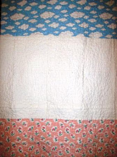
        I finished the Navaho Blues quilt and mailed it off. It is machine pieced, 54 in X 76 in. It is machine and hand quilted as well as tied. I did the hand quilting using crochet thread in a "big stitch" fashion.
        Then used the same crochet thread to "tie" some corners using a square knot (of course). You can see a few of the ties in this photo.

        Thursday, April 24, 2008

        Women in Medicine


        Updated 3/2017-- photos and links removed as many are no longer active

        Harriet Hall from the Science-Based Medicine blog wrote a piece on "Women in Medicine" recently. It is interesting reading, as I hope her book will be. I've ordered my copy.
        Something very interesting is happening in medicine. It’s happening slowly, quietly, and steadily, with no help from affirmative action programs.
        At the beginning of the 20th century about 5 percent of the doctors in the United States were women. In 1970, it was still only 7 percent. By 1998, 23 percent of all doctors were women, and today, women make up more than 50 percent of the medical student population. In 1968 only 1.2% of practicing dentists were women. By 2003, 17% of dentists were women, and 35% of dentists in new active private practice were female.

        Wednesday, April 23, 2008

        Dr Val Does Grand Rounds Her Way

        Updated 3/2017-- photos and all links removed as many no longer active.
         
        Grand Rounds is hosted by Dr Val this week -- "How do you feel about that?" [what color is your mood ring?]
        There are many approaches to summarizing submissions to Grand Rounds, and I have chosen one that has never (to my knowledge) been used before.
        That's right – I'm taking my inspiration from the limbic system, and have organized the posts according to the dominant emotion they elicit from readers. So without further ado, here's the Grand Rounds that will make you laugh, cry, stomp your feet, and become enlightened in the process.
        Next week Grand Rounds will be hosted by Jan Gurley, who has this special message:
        Grand Rounds in medicine often means a morgue-cold auditorium, a sea of starched white coats, and staccato squeaks from irritable chairs. Doc Gurley is hosting April 29th's Grand Rounds of the medical blogosphere with a more WWF-type approach: Grand Rounds Smack Down Week. Do you want to take on a behemoth topic with some chest-beating frenzy? Or just climb into the Internet ring wearing your most outrageous verbal-costume? Here's your chance to go for it.
        And don't forget to join us tomorrow night for The Doctor Anonymous Show at 8 pm CST. This week he will be without a guest, but the chat room will still be open and it is great fun!!!
        You can go here for Tips from Dr A for first time Blog Talk Radio listeners.

        Monday, April 21, 2008

        The Anophthalmic Syndrome

         Updated 3/2017-- photos and all links removed (except to my own posts) as many no longer active.

        See my previous post. It is the reason I went looking for more information. Until now I didn't know about the anophthalmic syndrome. As you can see from the articles below, it doesn't seem to be an area that garners much interest these days. Most of the articles (that I had easy access to) are pre-1990. The intro from the 4th referenced article puts the issue in perspective:
        The patient who has had the misfortune of losing an eye, be it secondary to disease or trauma, often has difficulty in getting the help he needs from the medical profession. Many ophthalmologists, being "eye surgeons," lose interest in a patient when the globe is gone. Many reconstructive surgeons hesitate to venture into this area which seems to be surrounded by a certain amount of mystique. Both may escape by referring the patient to an ocularist for continued care related to his appearance. The ocularist, who is not a medical doctor, often gains more practical knowledge of the problems of the anophthalmic state than does either the ophthalmologist or the reconstructive surgeon but, because he is unfamiliar with all possible reconstructive surgical techniques, he often fails to recommend reconstructive surgery when it is indicated. Lars M Vistnes, MD
        Within this post, all the issues will be discussed with the stipulation that there is a normal bony orbit. Only the soft tissue elements will be discussed. You may wish to read this description of the enucleation procedure.
        The anophthalmic syndrome consists of:
        • enophthalmos (eye appears sunken)
        • superior sulcus depression (upper eyelid crease is too deep compared to normal eye)
        • lower lid ptosis
        • upper lid ptosis
        Enophthalmos
        Deficits in the orbital volume contents and superior sulcus depression are recognized to be the cause of enophthalmos. Several caused have ben postulated (ref 1):
        • levator disinsertion
        • atrophy of orbital fat
        • loss of volume when the globe is removed
        • depression in the floor of the orbit due to an unrecognized fracture in the floor (rare)
        • malposition in the superior rectus muscle
        Replacement of lost volume can be done using a variety of materials -- autogenous bone, cartilage, dermis, glass beads, and silicone are some of the materials used. First, see what an experienced ocularist can do
        Superior Sulcus Depression
        As with, enophthalmos surgery should only be done if an experienced ocularist is unable to correct the problem. Correction of the two problems go hand-in-hand.
        Lower Lid Ptosis
        The pathomechanics is believed to be secondary to gravity acting with altered vectors of force on a prosthesis that is heavier than a normal eyeball. This appears to be independent of periorbital trauma.
         
        Correction of this deformity may be best done by use of a fascial sling (ref 2). There are two key features that need to be remembered in correcting lower eyelid ptosis.
        • The normal lower eyelid, with the eyeball in a horizontal gaze, has its upper border at the level of the lower margin of the limbus. The curvature of this border is not uniform. In its lateral third it assumes a more superior direction. To recreate this normal curvature the lower eyelid requires a higher positioning of the orbital rim burr hole than once thought.
        • A second consideration is the normal motor function of the inferior rectus. Through its connection into the capsulopalpebral ligament, the lower eyelid upon downward gaze is simultaneously pulled in a inferior direction. A static sling suspended between the medial canthal ligament and the orbital rim restricts this motion. Therefore, a static sling is most applicable in the case of an anophthalmic orbit in which a static lower eyelid does not interfere with vision.
        The fascia lata sling is described in the second reference article in great detail. I would like to relay the technical tips that Dr. Vistnes makes.
        1. The optimal surgical correction must begin with an ideal prosthesis. Such a prosthesis is made to fit the socket and does not attempt to compensate for enophthalmos or lower or upper eyelid ptosis.
        2. The width of the fascial strip is 2 mm. By pulling on a smaller area of the lower lid, directly below the lash margin, the lid can be positioned more precisely.
        3. The use of the Wright's needle allows the fascia to be passed under the space anterior to the tarsal plate. The needle can be positioned immediately beneath the lash margin, and the fascial strip will seat itself in this track without displacing itself inferiorly on the tarsal plate. Low placement of the sling can result in eversion of the lash border ("tumbling" of the lid into ectropion).
        4. Positioning the orbital burr hole at approximately 5-6 mm above the level of the lateral canthal tendon will not recreate the normal anatomy. The proper site can be chosen by following the curvature of the normal lower eyelid and marking the point where the curve intersects the orbital rim. An identical point on the anophthalmic orbital rim is then marked. This is the appropriated site for the orbital burr hole.
        Upper Lid Ptosis
        The cause will fall into one or more of three main categories:
        • the trauma which necessitated the enucleation
        • the surgeon (ie iatrogenic)
        • the surgery (ie the creation of an anatomical or pathomechanical situation that produces malfunction of a delicately balanced mechanism)

        There is a change in the size of the orbital support for the levator mechanism that occurs with enucleation. This, in most instances, is responsible for the upper lid ptosis. The "pivot" point of the levator muscle is lowered and more posterior than in a normal eye. Often this is corrected by experienced ocularist who add a superior sulcus "bridge" on the prosthesis thereby pushing the picot point of the muscle higher. (ref 4)
        Vistnes feels that the upper lid ptosis is a function of several factors:
        • the levator muscle tone and its adaptability
        • the tightness or laxness of the check ligaments
        • the size of the implants
        • the size of the prosthesis

        MANAGEMENT (according to Vistnes):
        When the implant is small and the prosthesis is large, and the degree of ptosis is moderate to severe -- correction may be obtained by a traditional levator shortening (Berke method).
        When the levator action is good over a prosthesis of average size and the ptosis is minimal, then a lid-shortening "ptosis correction" procedure may be used (Fasanella and Servat).
        The order in which the various operations are done in patients with the anophthalmic orbit syndrome (per Lars Vistnes MD)
        1) The volume deficit should be corrected first. In the cases of mild ptosis where an added mass (RTV silicone) is placed along the orbital floor, the pushing upward of the implant is often all that is required. This will also correct the enophthalmos and the superior sulcus depression.
        2) If lower lid ptosis is present, it should be corrected next (as a separate procedure). This correction will tend to push the prosthesis upward and may also correct the upper lid ptosis.
        3) Any ptosis of the upper lid should be corrected last -- and only after an experienced ocularist has been unable to correct it with a new prosthesis that is not out of proportion in appearance to the normal eye.
        I realize I have just begun to learn about the anophthalmic patient needs. I have not actually cared for any either in training or since. I hope this post will be of use to others who may be in a position to care for these patients. So if I made any mistakes, major or minor, please let me know so that I can correct this post. Thank you.
        Jarling Ocular Prosthetics, Inc --nice source of information on the actual prosthetics.
        The Artificial Eye Clinic -- another good source of information on the actual prosthetics.
        REFERENCES
        1. Correction of Enophthalmos and Superior Sulcus Depression in the Anophthalmic Orbit: A Long-Term Follow-Up; Plastic & Reconstructive Surgery. 79(3):331-338, March 1987; Sergott, Thomas J. M.D.; Vistnes, Lars M. M.D.
        2. Correction of Lower Eyelid Ptosis in the Anophthalmic Orbit: A Long-Term Follow-Up; Plastic & Reconstructive Surgery. 72(3):289-292, September 1983; Nolan, William B. III M.D.; Vistnes, Lars M. M.D.
        3. Blepharoplasty in Patients with an Anophthalmic Orbit; Plastic & Reconstructive Surgery. 59(5):670-674, May 1977; Horton, Charles E. M.D.; Graham, John K. M.D.
        4. Mechanism of Upper Lid Ptosis in the Anophthalmic Orbit; Plastic & Reconstructive Surgery. 58(5):539-545, November 1976; Vistnes, Lars M. M.D.
        5. Correction of Enophthalmos in the Anophthalmic Orbit; Plastic & Reconstructive Surgery. 51(5):545-554, May 1973; Iverson, Ronald E. M.D.; Vistnes, Lars M. M.D.; Siegel, Richard J. M.D
        6. Blepharoplasty, Upper Lid Ptosis Surgery; eMedicine Article, Jan 30, 2008; Jorge I de la Torre MD
        7. Correction of Superior Sulcus Deformity and Enophthalmos with Porous High-density Polyethylene Sheet in anophthalmic Patients; Korean Journal of Ophthalmology, 19(3):168-173, 2005; Byeung-hun Choi, MD; Sang-hyeok Lee, MD; Wha-sun Chung, MD (PDF file)
        8. Evaluation of the Anophthalmic Socket: It's Important to Understand the Management of Anophthalmic Patients and Recognize Complications; Review of Ophthalmology, Vol 13:09, Sept 5, 2006; Ann P Murchison MD and C Robert Bernardino MD


        A Singular View


        Updated 3/2017-- photos and all links removed as many no longer active.

        Back in March after writing the post on prosthetics, I received a package that contained a copy of the book, A Singular View, and a copy of the Journal of Ophthalmic Prosthetics (JOPPro) along with a note. Michael O Hughes, ocularist and senior editor of the JOPPro asked me to submit an article for a "focus" edition on esthetics that they are putting together for next year.
        My thought of an article (only four pages needed!!) would be expectations of the patient from the perspective of a plastic surgeon; which is very similar to the restorative work of an ocularist and a oculoplastic surgeon. Please keep in mind; ocular/facial prosthetics is very different from orthotics; which you may be more comfortable/familiar- due to your father.
        Facial disfigurements can hit a patient at the core of their security. Then again; some patients are very flip id about the loss. Regardless; the world of ocular prosthetics is surrounded by myths and many misconceptions. Maybe plastic surgery; for the average consumer is also.
        Michael Hughes
        I have no experience, either in training or in practice with patients who have lost an eye. I do not feel qualified to write such an important article. If there is anyone out there who can (perhaps David Khorram, MD who writes the blog marianaseye), please do so. Thank you.
        There is a lot of information at Mr Hughes website. The information covered includes ocular prostheses (photographs, the fabrication, and the history) and patient resources.
        As for the book, it is a great source of information on adjusting to monocular vision. It contains practical suggestions such as:
        • Lightly touch the pitcher of water/tea to the rim of the glass before pouring.
        • When choosing a seat at the dinner table, try to sit with your unaffected (remaining eye) side to the person you will be conversing.
        • Learn how to use perspective and relative movement to judge distance.
        • Don't give up the activities you enjoyed prior to your eye loss. Relearn how to do them with monocular vision. The books author was a pilot both before and after his injury left him with one eye.
        The book gives a nice explanation of how depth perception works and is changed by the loss of one eye. Depth perception involves 1) retinal disparity, 2) convergence, and 3) accommodation.
        Retinal disparity depends on an object being viewed with two eyes separated by several inches so that each eye is looking at the same target from a slightly different location at the same moment.
        Convergence has to do with the merging of these two images produced on the retinas. The effort by the eyes to bring the two images into exact correspondence produces a strain on each eye, and the experienced brain knows how to translate this into a measure of distance.
        Accommodation is a term for the automatic adjustment each eye makes to bring an object into focus. It is only effective for judging distances up to about six feet; thus it's likely to be the least useful of the three mechanisms. When you've lost an eye, however, it's the only one available to you, and we will cultivate it to its limit.
        There are many famous people who have had great careers and only monocular vision. Some examples are:
        • Peter Falk -- an actor well known for the detective Columbo. Surgeons had removed his right eye, along with a malignant tumor, when he was three years
        • Sandy Duncan -- an actress and dancer. In the 1970s, she was treated for a tumor behind her left eye, which damaged the optic nerve. She lost the sight in the eye.
        • Theodore Roosevelt -- 26th president of the United States. He lost his left eye in a boxing match with a naval officer
        • Sammy Davis Jr --singer/entertainer. He lost his left eye in an automobile accident prior to achieving stardom as an entertainer
        • Wiley Post, pioneering aviator who made the first solo circumnavigation of the globe with vision in only his right eye
        • Elizabeth Blackwell, the first woman to graduate from an American medical school lost an eye while in postgraduate school in France
        • James Stuckey, MD--not as famous, but did practice Plastic Surgery in Little Rock, Arkansas. He was very well thought of by his patients, his peers, and the nurses. He retired in the early 1990's and died almost 10 years ago. (Couldn't find a link to his obituary)

        Sunday, April 20, 2008

        Dr Schwab to host SurgeXperiences 120!!!

         Updated 3/2017-- photos and all links removed as many no longer active.

        The next edition of SurgeXperiences (120) will be hosted by Dr. Sid  Schwab from the northwest part of the United States near the Pudget Sound. He is a retired general surgeon whose blog is Surgeonsblog.  He is the author of the book "Cutting Remarks".  If you haven't read it, you should.
        He has been on the Dr Anonymous Blog Talk Radio show as both a guest and as a guest host. 
        Dr. Schwab's request for submissions:
        "Having complained previously about themed blog carnivals (while acknowledging that people have a perfect right to do it however they choose), I'm making no suggestions other than getting your entries in on time. Unless an unprecedented deluge leads to more than I can handle, I plan to link 'em all up. So feel free, and freed. And please: lower your expectations."
        SurgeXperiences is a blog carnival about surgical blogs. It is open to all (surgeon, nurse, anesthesia, patient, etc) who have a surgical blog or article to submit. The deadline for submissions is April 25th. Please submit your posts here.
        Here is the catalog of past surgXperiences editions for your reading pleasure. If you wish to host a future edition, please contact Jeffrey who runs the show here.

        P.S.
        Don't confuse "our" Dr Sidney M Schwab, surgeon, with Dr Sidney I Schwab, the neurologist.    Dr. Schwab are you related?

        Saturday, April 19, 2008

        My First Quilt Swap

        Updated 3/2017-- photos and all links (except to my own posts) removed as many no longer active.

        About a week and a half ago, I was invited by Kate to participate in a quilt swap. Kate lives in England (just outside of London). Her blog is Kate's Quilting (& other fibre arts) Blog. I "met" Kate when I won some Flying Geese quilt blocks from her back in September 2007. I made a baby quilt from them which you can see here. We now play Scrabulous together on Facebook. The wonders of blogging and the internet! I have enjoyed all the friends it has added to my life.
        So I will be participating in my first (Kate says they are addicting) quilt swap. Here are the rules:
        • Quilts should be 12" square minimum and 20" square maximum. They do not have to be square, of course.
        • All quilts should be finished (that is, quilted and bound) and should include a label on the reverse with your name, location, date and your partner's name, as well as the name of this swap.
        • Because these are small quilts intended for display rather than use, you may include surface embellishment techniques such as beading, embroidery, fabric painting and so on, but please ensure any technique you use is durable (being washable is not required).
        • Any technique or style is allowed, including whole cloth quilts or art quilts - and traditional quilts are welcome as well - so if you have a dislike of a particular style, be sure to note it when signing up.
        I have started playing around with "laced ribbons" using 1.5 inch squares. The pattern is a variation of the Double Irish Chain, using varied color to give the appearance of woven ribbons.
        As you can see from this list of the other participants, it is an international swap:

      • Amy (US)
      • Andrea (UK)
      • Ann A (US)
      • Ann J (France)
      • Ann R (US)
      • Anne B (UK)
      • Annette (UK)
      • Calico Cat (US)
      • Cat (US)
      • Cathi (Ireland)
      • Colene (US)
      • Colleen (US)
      • Dorothy (US)
      • Hedgehog (Finland)
      • John (US)
      • Jovita (Lithuania)
      • Kate (UK) - hostess
      • Liliane (Luxembourg)
      • Linda (US)
      • Lisa (US)
      • Lisa Jo (US)
      • Lorraine (Australia)
      • Loulee (UK)
      • Lynda B (Australia)
      • Lynda M (UK)
      • Margaret (US)
      • Paula (US)
      • Sarah (US)
      • Sequana (US)
      • Shelly (US)
      • Sherry (Ireland)
      • Solidia (US)
      • Sophie (US)
      • Stephanie (UK)
      • Teresa (UK)
      • Toni (US)
      • Tracy (Australia)
      • Wil (Netherlands)
      • Becca (US)
      • Dawn (Turkey
      • Friday, April 18, 2008

        Breast Reconstruction Webcast


         Updated 3/2017-- photos/videos and all links (except to my own posts) removed as many no longer active.

        Yale Surgery Update Blog featured a copy of webcasts recently that were nominated for awards. The one of interest to me was Breast Cancer: Reconstruction Immediately Following Radical Mastectomy from Thomas Jefferson University Hospital, originally broadcast October 18, 2007. It shows breast reconstruction using a latissimus dorsi flap with expander. It is worthy of the award and is well worth watching.
        You may also wish to review my past post on Breast Reconstruction--Part I and Part II

        Thursday, April 17, 2008

        Cat Bites

         Updated 3/2017-- photos and all links (except to my own posts) removed as many no longer active.

        A blog friend was recently bitten by her cat while she was giving said cat a bath. This wouldn't in most of us be much of an issue, but this blog friend is immunosuppressed. This post is for her.
        Animal bites are not uncommon occurrences. The ones seen in emergency centers represent only a small percentage of all bite victims. The majority of reported animal bites are dog bites (80-90%). Cat bites make up only approximately 10%. The rest are bites from miscellaneous animals and rodents.
        Most animal bites occur on the extremities, but the head and neck region is also often affected. The head and neck region is injured in 6-20% of persons who sustain cat bites. Children are injured more frequently in the head and neck region than adults.
        The incidence of infection transmission is quite low. The risk of rabies is probably the best reason for investigating animal bite injuries. Most cat bites are from pets rather than strays, so the rabies vaccination history should be available.

        Medical Care
        • IF the skin is only contused and not broken, then simply cleanse the skin thoroughly with soap and water. There is no reason to seek further care unless signs of infection (see below) occurs over the next few days.
        • IF the skin is penetrated, copious irrigation is warranted, in addition to thorough cleansing. See your medical doctor or go to the emergency department, especially if you are immunosuppressed or your tetanus is not up-to-date or there is more to the injury (lacerations, continued bleeding, etc).
        • Basic wound management is the key. Treatment may include debridement, antibiotic therapy, supportive care, and, possibly, primary suturing or hospitalization with operative debridement. Only 1-3% of all bite injuries (dog, cat, etc)require hospitalization for surgical debridement and intravenous antibiotics.
        • Tetanus toxoid is administered. The rabies status of the animal will be investigated.
        • The force of a domestic cat's bite does not match that of a dog. Its sharp teeth may cause a puncture wound into which bacterial organisms are inoculated. The risk of infection is compounded by the feline habit of paw licking, which may contaminate their claws with oral flora. The risk of infection is higher following a cat bite than a dog bite. Also, cat bites carry the risk of causing catscratch fever with resultant adenopathy. This is usually self-limited except in immunosuppressed patients.
        • Signs of infections are typical and include redness(rubor), pain (dolor), heat (calor), and swelling (edema) of the tissues. Purulent discharge from the wound is another good indicator of infection. Signs of infections may appear 24-72 hours following the bite.
        Antibiotic Use in Catscratch Disease
        Patients with mild-to-moderate catscratch disease should not receive antibiotics.
        For patients with extensive symptomatic lymphadenopathy, azithromycin is recommended.
        All immunocompromised patients with catscratch disease, however, should be treated with antibiotics.
        Bartonella is an intracellular bacterium and has poor response to penicillin derivatives in vivo despite susceptibility in vitro. A single treatment for all Bartonella-related diseases has not been identified, so treatment must be adapted for specific situations.
        Bartonella infections are generally treated with macrolides, tetracyclines, aminoglycosides, or chloramphenicol.
        Duration of therapy is commonly at least 3 weeks. Patients should be monitored for evidence of response and drug toxicity. Because these infections often fail to respond to therapy or patients experience relapse later, switching to antibiotics from other classes (eg, erythromycin, clarithromycin, azithromycin, trimethoprim and sulfamethoxazole, or ciprofloxacin) may be needed. Gentamicin may also be effective.

        For retinitis, a combination of doxycycline and rifampin for 4-6 weeks is recommended.
        For bacillary angiomatosis and peliosis hepatis, long courses of erythromycin or doxycycline are recommended for 3 and 4 months, respectively.
        In known Bartonella endocarditis, a combination of doxycycline for 6 weeks with gentamicin 3 mg/kg/d IV for 14 days is recommended.
        Some patients, usually who are immunocompromised, develop a Jarisch-Herxheimer–like reaction shortly after receiving antibiotic therapy.
        You may also wish to check out my previous post on "Preventing Dog Bites".
        REFERENCES
        Catscratch Disease; eMedicine Article, August 15, 2006; Roseanne Ressner DO, Lynn Horvath MD
        Animal Bites; eMedicine Article, August 1, 2006; Suzanne Galli MD, Philip Miller MD
        Bartonellosis; eMedicine Article, October 31, 2007; Kassem Hammoud MD, Daniel Hinthorn MD
        Cat Scratch Disease: Bartonellosis; Katharine Hillestad, DVM; Drs. Foster & Smith Veterinary Services Department
        From Cat Scratch Disease to Endocarditis, the Possible Natural History of Bartonella henselae Infection; BMC Infect Dis 2007; 7:30; Frédérique Gouriet, Hubert Lepidi, Gilbert Habib, Frédéric Collart, and Didier Raoult

        Wednesday, April 16, 2008

        Update on Marathon Death

        Updated 3/2017-- photos and all links (except to my own posts) removed as many no longer active.

        The Little Rock Marathon took place on March 2nd. We had a runner, Adam Nickel, collapse after finishing the race in 3:02:26. He was not known to have any medical problems. He finished 18th overall in the marathon.
        Autopsy Provides Answers, but little solace
        Autopsy results from the Arkansas Crime Lab, released Monday, indicate that Nickel had a vascular disease known as “multifocal small coronary artery fibromuscular dysplasia,” which means he had small heart arteries that restricted the flow of blood. The small arteries were near a critical part of the heart that regulates electrical impulses.
        That, combined with abnormal electrolyte percentage levels that many distance runners experience, including raised potassium percentage levels, led to a significant heart arrhythmia that proved fatal.
        Dr. Charles Kokes, the crime lab’s chief medical examiner, said an electrocardiogram could have detected Nickel’s condition, but a physical alone would not.
        Many runners might have Nickel’s condition, not know it and still feel fine, Kokes said. In Nickel’s case, the fact that the affected arteries were so near nodes in the heart that control electrical impulses led to the fatal event.


        Dr. Anonymous Blog Talk Radio


        Updated 3/2017-- photos and all links removed as many no longer active.
        Join us tomorrow night for The Doctor Anonymous Show at 8 pm CST. His guest will be Vijay from Scan Man's Notes who lives and works (radiologist) in India. He'll be calling in live.  It'll actually be Friday morning for him. 
        In addition to just listening to the show, there is a chat room that we "listeners" gather in. It is great fun! Hope you will join us.

        Tuesday, April 15, 2008

        It Costs How Much?

        Do you remember this splint that I was told to wear (and did for a week) when I had acute olecranon bursitis? Well, I got my bill recently and was shocked by how expensive the splint was! If I had been told how much it was going to be, I swear I would have left without it. I would have used the "soft" elbow pad and made myself a plaster splint to protect my elbow from being bumped for that week. Here's the breakdown of the medical bill. For some reason, all of the charges had been billed to insurance EXCEPT the splint charge. I asked that it be submitted.

        ServiceDescription of ServiceChargeInsurance AllowedPatient to Pay
        99204Office/New Patient$229.02$140.38$140.38
        73070X-Ray Exam$73.92$33.12$33.12
        97760Orthotic (OT)Management$40.00$36.00$36.00
        L3763Rigid Elbow Splint$773.33??????

        Monday, April 14, 2008

        Where are you from?

        What do Scotland, Damascus, London, Berlin, Sweden, Moscow, England, and Jerusalem have in common?

        They are all towns in Arkansas (USA). Seriously.

        I say this to give some background for my story. I had a patient come into my office whose last name happened to be Bates. "Where is does your family come from?"

        "England"

        "Well, my family came from Cotton Plant/Brinkley part of the state. So we might be related a few generations back."

        A blank look on there face. "We moved from England to Canada when I was little. Later to Washington state before moving to Arkansas."

        Now (mental head slap) I got it. They didn't even realize there was a town called England in Arkansas. It was now my turn to explain.

        Sunday, April 13, 2008

        SurgeXperiences 119 is Up

        Updated 3/2017-- photos and all links removed as many no longer active.

        SurgeXperiences 119, hosted by Dr. David Khorram from the Saipan, is up. He is an eye surgeon who's blog is MarianasEye. He is also the author of the book "World Peace, a Blind Wife, and Gecko Tails".
        Dr Khorram title this edition of the surgery carnival: Funny Operating Room Moments. There are some good/great posts, but he makes it better with his commentary.
        "I was filming a nipple reconstruction..." Thus begins a "Tale of Two... "um, no, I'm not going to say it... a tale by Norwegian surgical videographer, The Sterile Eye, who presents Yo mammae! That sentence is sure to become a classic in medical blogging literature. I give Sterile Eye the "Call me Ishmael" Award (which I just created) for week's best opening sentence of a Carnival post.
        SurgeXperiences is a blog carnival about surgical blogs. It is open to all (surgeon, nurse, anesthesia, patient, etc) who have a surgical blog or article to submit. The next edition will be hosted by Dr Sid Schwab, Surgeonsblog, on April 27th. The deadline for submissions will be April 25th. Please submit your posts here.
        Here is the catalog of past surgXperiences editions for your reading pleasure. If you wish to host a future edition, please contact Jeffrey who runs the show here.

        Saturday, April 12, 2008

        Oh Sunny Day--How Sweet!

        Today was absolutely beautiful! Especially wonderful after so much stormy weather here in the past few months. There are many areas in Arkansas where the flooding persists due to the continued rain. But today was sunny and it was sweet! I spent several hours outside just enjoying the day. So apparently did the small green lizard on our fence.
         
        I started another baby quilt today. I'm using the Houndtooth Scrap Patch pattern in some bright colors.
        Each block will be 10 inches sq. I'm going to do the stashing in blue. I haven't decided about the border yet. Here is one of the finished blocks. Bright and sunny, just like the day.

        Friday, April 11, 2008

        Square-in-a-Square Baby Quilt

        Here is a baby quilt I made for a circulating nurse that I adore. She had two boys and wanted a girl. She wouldn't let her Ob-Gyn tell her the sex of the baby. I found this cowgirl fabric, but resisted the urge to use more pink than I did in the quilt. I used some blue, green, and pink just in case. She got her girl though.

        The pattern is a square-in-a-square. The quilt is 40 in X 51 in. It is machine pieced and quilted. You can see some of the quilting in these two detail pictures.

        Thursday, April 10, 2008

        Sutures and Wound Care

         Updated 3/2017-- photos and all links removed as many no longer active.

        How do you have patients take care of their incisions? I usually tell them that it is okay to shower (no tub baths) the day after surgery. I encourage the use of soap and water. I think it helps keep the incision clean. It also encourages them to move. There are exceptions to this--skin grafts, hand surgeries, etc--where they are told to keep the dressing clean and dry.
        I read an article recently regarding this basic premise of postop wound care and how it affects the sutures used for wound closure. The article was by Raghu Athre, M.D., of the Department of Otolaryngology — Head and Neck Surgery at Southwestern Medical Center, University of Texas, Dallas. The study was suggested by an observation in postsurgical patients after they had undergone surgical treatment for head and/or neck cancer.
        Owing to the equivalent scar profile of absorbable suture and permanent suture, and the ability of the surgeon to conserve time and minimize patient anxiety by using absorbable suture, most surgical patients have the superficial layer of their surgical wounds closed with 5-0 fast-absorbing gut suture. The main criterion for this decision is surgeon preference. At 1-week follow-up, a subset of these patients show dehiscence of the most superficial layer of skin closure. The superficial epidermis pulls apart, and no evidence of the fast-absorbing gut sutures can be found. The separated wound closes by secondary intention over the ensuing 3 to 4 weeks without significant complications. Extensive questioning of these patients reveals that most of them used hydrogen peroxide to clean their wounds postoperatively. The question of whether hydrogen peroxide affects the tensile strength of fast-absorbing gut sutures by increasing its degradation rate became the focus of this study.
        So he and his team conducted a laboratory study comparing the tensile strength of nylon, polypropylene and fast-absorbing gut sutures. In the study, each suture type was treated with either three percent hydrogen peroxide or water for a period of five days, emulating wound care following a surgery. Then an In-Spec 2200 bench-top tester was used to assess the maximum tension that the sutures could bear prior to breaking. The results:
        On preliminary visual examination, nylon and polypropylene sutures did not appear to be affected by either water or hydrogen peroxide. Both groups of suture samples retained their shape, color, and general feel when handled. The fast-absorbing gut suture, however, was different. The fast-absorbing gut suture subjected to water did not appear to be affected compared with control suture. The fast-absorbing gut suture subjected to hydrogen peroxide rinses completely disintegrated during handling. It could not hold any tension at all, and in 1 case, the hydrogen peroxide–treated suture had completely degraded; the only thing left behind was the needle.
        Creating a cosmetically appealing scar that does not affect form and function is one of the important endpoints of a surgical procedure. Complications such as wound infection, wound dehiscence, hypertrophic scarring and contractures may all result from improper closure techniques, improper wound-care regimens — as well as patient factors such as nutritional status and medical comorbidities.

        FUNDAMENTAL CHANGE IN THOUGHT NEEDED
        Many standard superficial wound-care regimens include water, soapy water, antibiotic ointments (neosporin, bacitracin, bactroban, etc), hydrogen peroxide and Dakin's solution, or any combination of these. Patients are very likely to use hydrogen peroxide unless told not to do so. We surgeons need to give specific instructions to patients for home care. [This could also be said about any physician who repairs lacerations, etc.] We may want to strongly advise them to avoid use hydrogen peroxide, and to select saline or soap and water for wound cleaning instead.
        The results of this study underscore the importance of the agents used in wound care following a surgery. Hydrogen peroxide should be avoided as a superficial wound-care regimen where fast-absorbing gut sutures are used.

        REFERENCE
        Athre RS, Park J, Leach JL. The effect of a hydrogen peroxide wound care regimen on tensile strength of suture. Arch Facial Plast Surg. 2007;9:281-284.
        Dr RS Athre interviewed by Cosmetic Surgery Times on his study; April 2008 Issue

        Wednesday, April 9, 2008

        Dr. Anonymous' BlogTalk Radio Show


         Updated 3/2017-- photos and all links removed as many no longer active.

        Join us tomorrow night for The Doctor Anonymous Show at 8 pm CST. His guest will be none other than Bongi from Other Things Amanzi who lives and works (general surgeon) in South Africa. He'll be calling in live (about 4am where he's lives). In addition, Dr Sid Schwab will be co-hosting with Dr. Anonymous. Also, mark your calendar for next week, April 17th when Vijay from Scan Man's Notes will be his guest. Vijay is a radiologist in India.
        In addition to just listening to the show, there is a chat room that we "listeners" gather in. It is great fun! Hope you will join us.

        Tuesday, April 8, 2008

        Druthers

         Updated 3/2017-- photos and all links (except to my own posts) removed as many no longer active.

        What a nice word--druthers. It is a noun, pronounced DRUTH-uhrz. It is a contraction of "d rather" as in I would rather. And that is it's meaning -- one's own way; preference.
        Every operating room I have ever worked in has "preference cards" for each surgeon. Very often for each procedure that surgeon does. The card covers the surgeon's glove size (and sometimes type, powderless, etc), preferred skin prep, drapes, patient position, etc. Often the music preference is included on the card. It includes a list of instruments and sutures/needles. It includes the "preferred" dressings for the surgical incision.
        I druther have the patient's face prepped with Technicare Surgical Scrub than Betadine.
        I druther have Adson's with teeth (fine) than Brown's forceps.
        I druther have fine needle holders, smooth like Webster's.
        I druther not use towel clamps, but like having them just in case.
        I druther have 94.1 FM. You know, the station that my sister and niece won the New Zealand trip from.
        I druther have no foley today. I think we'll be less than 3 hours and she's young.........
        I druther have the suture in the room and not open it until we need it. We won't waste as much that way.
        I druther patient's not have any postop nausea.
        I druther have ........

        Along theses lines, if there are any residents or fellows who read this blog I would encourage you to ask for copies of your "preferred" staff's preference lists for a few major and minor surgeries. It will be most helpful to you when you move out into your practice. The new hospital(s) will appreciate it as they get used to your routine. When I left my residency a wonderful scrub nurse did that for me. Back then the cards were hand-written, now most places should be able to print it out from their computers.

        Monday, April 7, 2008

        University of Arkansas Physics Centennial

         Updated 3/2017-- all links removed as many no longer active.


        I am back from attending the UA Physics Centennial. It was wonderful. Everyone was so happy to be back, telling stories of their time at UA and their lives since. Many have remained in physics --research, teaching, industry. Many others are doctors, lawyers, even a wine maker.

        The lecture given by Dr. Wolfgang Ketterle was clear and easy to follow. The title of the lecture is "New Forms of Quantum Matter Near Absolute Zero temperature". It was nice to see children and teens there along with the rest of us.

        Part of the celebration was the unveiling of a George Rhoads' audiokinetic sculpture that is now in the entry way of the Physics Building. I wish I could share the actual motion with you, but here is a picture of it.


        This photo is of all the past and present professors who were there. I know I can't get them all in order. The one in the blue/red coat in front is Dr. Raymond Hughes. The one just to the left is Dr Rajendra Gupta. The one in the center, brown leather coat is Ken Vickers (microEP program director). Just to the right is Dr Otto (Bud) Zinke. The one with the red shirt is Dr Steven Day. Just behind him and to the left is Dr Greg Salamo.

        This photo is of Dr Richard Anderson
        and his students who attended.


        While I was gone, Arkansas had 6 tornados (gallary of photos) come through the state. Several did damage near my home. I am fortunate that I was able to return to a house that was not harmed by the storms, neighbors who checked on things for me, a dog who was glad for me to pick him up from the vets', and sunshine. My husband is currently working out-of-town so wasn't there to greet me. Still it was a nice to be home.

        If you are interested in the history of Physics you can read Physics at Arkansas by Paul Sharrah by linking here.

        Sunday, April 6, 2008

        SurgeXperiences 119 Call for Submissions

         Updated 3/2017--all links removed as many no longer active.

        The next edition of SurgeXperiences (119) will be hosted by Dr. David Khorram from the Saipan. He is an eye surgeon. His blog is MarianasEye.
        Dr Khorram is asking for
        So, medical people around the world, send me your funny stories. They don't have to be from the operating room per se (or more aptly the "operating theatre" as our British influenced colleagues would say). Write about any funny experience that occurred in the vicinity of a knife.
        SurgeXperiences is a blog carnival about surgical blogs. It is open to all (surgeon, nurse, anesthesia, patient, etc) who have a surgical blog or article to submit. The deadline for submissions is April 11th. Please submit your posts here.
        Here is the catalog of past surgXperiences editions for your reading pleasure. If you wish to host a future edition, please contact Jeffrey who runs the show here.

        Friday, April 4, 2008

        Story Book Baby Quilt

        Another niece is having a second baby this spring. I took a "fabric book" panel and used the pages as blocks. Then used strips in a woven pattern to put them together. It is 38 in X 52 in. The quilt is machine pieced and quilted.
        A detail view of some of the blocks.
        The back is made of three flannels pieced together.
        I didn't have enough of any of them seperately, but liked the result.

        Thursday, April 3, 2008

        Facial Duplication

         Updated 3/2017-- photos and all links removed as many no longer active.

        Facial duplication is also known as diprosopus. The word "diprosopus" comes from the Greek "di-," two and "prosopon," face and means "two-faced".
        Two-faced conjoined twins have almost complete fusion of their bodies with one set of limbs with part or all of the face duplicated. The condition usually results in stillbirth, but not always as seen in this recent news report of this little girl born March 11, 2008.

        Dr. Kotrikova (1st reference) writes that the "first description of a double face was mentioned in the chapter The Miracles of the Far East from Jacob van Maerlant's manuscript The History of Troyes dated approximately 1270. In the sixteenth century, Jacob Rueff, a barber surgeon, published in his Manual for Midwives woodcarvings of infants with lesser degrees of twinning, including diprosopus."
        However, a New York Times article by William Honan published in August 2000 states that the ancient two-faced Mexican figurines known as the "pretty ladies of Tlatilco" are now thought to represent diprosopus. It is believed these early Mesoamerican figurines may, in fact, be "the oldest scientifically medical images in world history." Sculpted over a period of 500 years beginning in 1200 BC, these little statues show diprosopus with two faces side by side. This degree of accuracy in documenting the anatomical and pathological features of a human head does not appear for another two millennia until the 16th-century studies of Hans Baldung Grien and Andreas Vesalius who did anatomical drawings based on dissected cadavers.

        Children with craniofacial abnormalities such as diprosopus are characterized by a single body and a variety of duplications of craniofacial structures. These children may have four separate eyes (diprosopus tetraophthalmus) or fusion of the medial eyes. Two, three, or four ears may be present. Some have only partial or complete doubling of the nose (diprosopus dirrhinus).
        The prevalence of conjoined twins (Siamese twins) is reported to be between one in 2800 and one in 200,000 deliveries. The prevalence of diprosopus is reported to be one in 15,000,000.
        Conjoined twins are classified according to their symmetry, site of fusion, and degree of duplication. The most frequent type of conjoined twins is thoracopagus (32.7 percent) and the rarest type is diprosopus (0.4 percent).
        Cardiac malformations (ventral septal defect, transposition of major arteries) represent the most common congenital abnormalities. The esophagus, stomach, and intestines may be duplicated. Where there are more severe associated anomalies, the prognosis is poor.
        Hat tip to Dr Kevin for the news article link.
        REFERENCES
        Operative Correction and Follow-Up of Craniofacial Duplication; Plastic & Reconstructive Surgery. 119(3):985-991, March 2007; Kotrikova, Bibiana M.D., D.D.S.; Hassfeld, Stefan M.D., D.D.S.; Steiner, Hans H. M.D., Ph.D.; Hahnel, Stefan M.D., Ph.D.; Krempien, Robert M.D., Ph.D.; Muhling, Joachim M.D., D.D.S.
        Facial duplication. The unique case of Antonio. J. Maxillo-facial Surg; Plastic & Reconstructive Surgery. 64(4):571, October 1979; Krupp, Serge
        Conjoined Twins; eMedicine Article, Jan 22, 2008; Khalid Kamal MD and others

        Wednesday, April 2, 2008

        She's a Beautiful Girl!

        The first time I met this girl, she sat in her mother's lap and tried to burrow into her. The girl was so shy, it was tough to get her to let me see the "thing" that had brought her to my office. The "thing" was a small (0.8 X 1.6 cm) congenital nevus on her face.
        The girl was preschool age. Mom really wanted the nevus removed. She was afraid it would be a "problem as it has gotten a little bigger". I tried to tell her that yes, congenital nevi can change into skin cancers, but it would be safe to simply watch her daughter's until she was older. At her current age we would need to go to operating room and use general anesthesia. If we waited until she was older, we would be able to do this one in the office with local. I emphasized that she would be trading the mole for a scar. A scar that hopefully would be thin, flat, and fade over time. I insisted that she go home and discuss all this with her husband and the little girl.
        We did the surgery. It (fortunately) was textbook perfect -- anesthesia and the mole excision. The versed was wonderful! The family was very happy with her/their care pre-and post-op at the surgery center.
        The day she returned for follow-up, she burst through the office door ahead of everyone (mom, grandma, and little sister) and greeted me with a hugh smile. I, in turn, smiled and ask her to come on back with me. No hesitation. Practically danced her way back. I ask her to let me sit her up on the exam table. Sure, no problem. No need for mom's lap this visit. I told her I just wanted to look, there were no stitches to take out. She is still smiling. "I'm a beautiful girl"
        Her mom proceeds to tell me that her daughter has been telling everyone "A good doctor made me beautiful!"
        I told her "Thank you, but you were beautiful already."
        Still she made my day.
        It always amazes me how some small thing like a mole/birth mark removal can bring a child out of their shell. Absolutely amazing!
        And yes she is a beautiful girl!

        Tuesday, April 1, 2008

        Grand Rounds in Six Parts, no that's Seven

        Updated 3/2017--  all links (except to my own posts) removed as many no longer active.

        I think I have made it to all the hosts.....
        Grand Rounds started at GruntDoc this week...
        He lost steam and asked Dr. Val to pick up...
        She thought the submissions were beneath her and asked David E Williams to continue...
        He was humbled by the excellent writing and asked Medgadget to step in.  They did an admirable job and finished by announcing "one milestone to report:   .....Artificial Intelligence has come far enough to allow virtual blog carnival hosting.
        Behold, Dr. Anonymous, an entirely official construct."
        He then turns it over to Dr. Rob and Musings of A Distracible Mind.
        But wait, that's not all!  Dr Rob sends us over to  Kim at Emergiblog!!! 
         
        Good work by all of you!  Lots of great fun on this April's Fool Day!  Thanks for including my post, Dr Val.
        Next week's  host will be Dr. Wes, cardiologist extraordinaire!

        Trigeminal Trophic Syndrome

         Updated 3/2017-- photos and all links removed as many no longer active.

        I don't recall ever reading about this syndrome or seeing a patient with it. The picture of the patient in the January 2008 (first reference) case presentation caught my eye. I was drawn in and wanted to know more about trigeminal trophic syndrome (TTS). I thought I would share it with you.
        It seems that TTS was first described by Wallenberg in 1901. It was described as an uncommon clinical entity in which cutaneous trophic ulceration develops within trigeminal dermatomes. The trigeminal nerve (CN V) is primarily a sensory nerve and is responsible for sensation in the face.
        TTS is a rare consequence of damage to the trigeminal nerve itself or its central sensory connection. The average period from the time of trigeminal nerve injury to the onset of the ulcer varies from several weeks to many years. Patients complain of picking, rubbing, or scratching sensations in the affected areas. These sensations are attributed to hypoesthesia, paraesthesia, or pain following the nerve damage. Conditions that can affect the trigeminal nerve include brainstem cerebrovascular disease, diabetes, tabes, syringomyelia, and postencephalopathic parkinsonism. It can also occur following surgical management of trigeminal neuralgia.
        The diagnosis is clinical. It is suggested when loss of sensation occurs in association with unilateral facial ulceration, especially the nasal ala. Patients tend to complain of paresthesias and painless traumatization.
        The differential diagnosis includes basal cell carcinoma, infectious diseases (tertiary syphilis, recurrent herpes simplex, leishmaniasis, cutaneous tuberculosis, or leprosy) or systemic disease (Wegener granulomatosis, Horton disease). Similar lesions may occur with factitial ulcers, but the psychiatric context is different.
        The characteristic lesion is a painless, sickle-shaped lesion involving the nasal ala. The margins are free. The ulcer base has scant crust. Other areas that may be included with these ulcers include the frontal scalp, ear, cheek, temple, and cornea. Once the ulcers appear, they are very persistent. Neurological examination will reveal decreased sensation of pain and thermal perception over the trigeminal area (very important for making the diagnosis).
        Management of trigeminal trophic syndrome is often difficult. Application of occlusive dressings can reduce trauma of the affected area. Hydrocolloid dressings help the ulcers heal. Treatment regimens involving amitriptyline and diazepam in addition to chlorpromazine and pimozide have met with some success. Carbamazepine is an effective therapy in trigeminal neuralgia and atypical facial pain because these agents may influence both the paresthesias and behavioral factors in this syndrome.
        Surgical repair is usually unsuccessful. Surgical repair may be worthwhile, however, if tissue with functional innervation and its own blood supply is used.
        Didn't share the picture from the first reference (the one that caught my attention). In it, the entire ala of the nose and more was missing. It is similar to the one that can be found here (be cautioned -- it's graphic).
        This is not a joke post. I love jokes, but am not good at telling them. I say this as it is April Fool's Day! There are others who are good at pulling off good jokes -- check here and here.
        REFERENCES
        Trigeminal Trophic Syndrome: Diagnosis and Management Difficulties; Plastic & Reconstructive Surgery. 121(1):1e-3e, January 2008; Bhatti, Ahmad F. F.R.C.S.I., A.F.R.C.S.I.; Soggiu, Daniela M.D.; Orlando, Antonio F.R.C.S., Dip.E.B.O.P.R.A.S.
        Skin Ulceration in Trigeminal Trophic Syndrome: Report of a Lesion Occurring 22 Years Later; Plastic & Reconstructive Surgery. 116(6):1814-1815, November 2005; Yiacoumettis, Andreas M.D., Ph.D.; Vlachos, Spiros M.D., Ph.D.
        Trigeminal Trophic Syndrome: A Report of Two Cases with Review of Literature; Dermatology Online Journal 9(5):26; A Elloumi-Jellouli, S Ben Ammar, S Fenniche, M Zghal, H Marrak, and I Mokhtar
        Trigeminal Trophic Syndrome; Mayo Clin Proc 1997; 72:543-545; Charles H Dicken MD