

Sunday, July 26, 2009

SurgeXperiences 302

Updated 3/2-17-- all links removed as many no longer active and it was easier than checking each one.
 
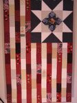
Once again I it brings me great joy to expose you to surgery (via blog posts rather than actual) and the many experiences that go along with it.   I hope you find them interesting. (this -- photo credit—makes me want to quilt it-- photo removed 3/2017)

Let’s begin with this one by a non-surgeon who finds scars as fascinating as I do.  Dr Charles’ post Cicatrix is much more elegant than I could ever write about scars, but captures my feeling wonderfully.  Each scar has a story attached to it.  We all have one scar in common – the umbilicus – which tells of our attachment to our mothers.
“It looks like it’s healing well.” I told her. I didn’t know if it would be appropriate or not, but I decided to say it anyway. “Your scar is a story. I hope you can own it. Don’t ever be ashamed by it. It’s a testament to your strength, a mark of your courage.”
Dr Charles was inspired by this article in the NYTimes “Our Scars Tell the Stories of Our Lives” by Dana Jennings, as was Tara Parker-Pope “The Power of Scars.”  The comments attached to Parker-Pope’s are a very interesting  discussion on scars and their stories.
…………………………………..
Moving on to the gratitude (or lack thereof) associated with the outcome of surgery, Bongi tells us about a patient with acalculous cholecystitis.
…….some time after this the neurosurgeon got a call from the family doctor from their home country. he was indignant. he wanted to know why the gallbladder had been removed in such a young male, a group that usually does not have gallbladder problems and that in the absence of gallstones. he felt it was totally unnecessary and demanded an explanation……..
……………………………………
QuietusLeo, the Sandman, tells us of true gratitude showed by a patient’s family in the form of a gift after taking care of a “12 year old boy needed ultrasound guided drainage of perforated appendicitis with abscess.”   The father sent a touching note along with the gift which you should go read.  The child (the patient) was less grateful, but we’ll chalk that up to his age.
I called the family to thank them for their generous gift. The son answered the phone. When I identified myself he said that I had done a poor job because he didn't see the James Bond movie. I apologized and assured him that it was much more important to be healthy again.
He then passed the phone to his father. I thanked him for the gift. He said it was only a small token of appreciation. I assured him that it was much more than that.
……………………………………………
Can you imagine the story that goes with this?! (photo removed 3/2017)  Check it out over at M.D.O.D. (photo credit)


T, Notes of an Anesthesoboist, back from her vacation in France tells us about a call to the ER -- “you know it’s bad when”
One day I was giving someone a lunch break and wheeling her patient to the O.R. with the circulating nurse when we heard over the P.A. system,
"Any available surgeon stat to the emergency room. Any available surgeon to E.R. stat."
That stopped us in our tracks for a second. The nurse and I exchanged a look.
"Sounds pretty bad," I said.
………………………………………..
Buckeye Surgeon talks about tough decisions as he gets ready to put in an access port for chemotherapy.
The other day I picked up the chart of a lady who needed a port and the first thing I noticed was that she was 92 years old. I must admit, my first thought was: what the hell are we doing here? She had metastatic breast cancer with lesions seen in her lungs and liver. I was all ready to march into the room and have an honest, heart to heart talk with the patient and the family about futile care and cost effectiveness etc etc.
But I composed myself. Every situation is different. I asked questions. I listened.

Bongi tells us about the difficulties of just walking away when nothing can be done surgically.   He doesn’t do as his “teacher” taught.  Go read the entire post.
"now why did i just walk away?" he asked. we all gave the usual blank stares. "because there is nothing we can do for her." he said with a chuckle. those of us who needed to be in his good books gave the obligatory half hearted laugh. i could just manage a smile that i think came out more as a grimace.
…………………………………………….
Orac, Respectful Insolence, writes about being faced with a different kind of tough decision in “iron surgeon?”  [first posted in July 2007 and reposted recently]
The other day, Sid Schwab, surgeon blogger extraordinaire, brought up a question that, I'm guessing, most nonsurgeons wonder about from time to time when contemplating how it is that we surgeons do what we do.
What about bathroom breaks?
…………….It happened to me only once, but it provided a serious dilemma. What do I do? I'm captain of the ship of the O.R., so to speak. The entire team depends on me. The patient depends on me.
And that's the key to making the correct decision……..
……………………………………………
From WhiteCoat’s Call Room -- What’s The Diagnosis #4 – this 13 year old boy will certainly have a story to go with his scar! 

Uveal Blues tells us about Better Vision, With a Telescope Inside the Eye for people with irreversible, advanced macular degeneration.  Check out the post for photos.
……………………………………….
Vijay, Scan Man’s Notes, gives us a quiz using “three x-rays”
A small mental exercise for medical bloggers.
See the following three portable (bedside) chest radiographs that were taken in an ICU setting. They are in sequence.
See if you can guess the story that they tell.
…………………………………………
What do you do when you can’t get good IV access?  Check out Dr Michelle Lin’s post  Sneak Peak "Trick of the Trade": IO line for failed IV access.  There’s even a video showing how to do the IO (intraosseous) access in adults.
Adult intraosseous needles are coming more into favor in the United States, although they have been part of standard practice in the military and Europe. Various commercial devices exist. The one we have at SF General is the EZ IO Needle. (I have no financial ties with the company.) Needle placement is surprisingly easy and takes less than 10 seconds, especially if you channel your inner Home Depot self in using the power drill.
……………………………………….
Orac does a much better job than I did discussing “Overdiagnosis of breast cancer due to mammography” and it’s implications.  It’s a must read for all of us.

M.D.O.D talks about a surgical tragedy in the recent news in two posts – here and here where he attempts to explain how the tragedy might have happened.
Surgeon hands intern trochar. Intern, stepping up on small stool puts all her weight behind it and hubs it. Descending aorta pierced, Surgeon apoplectic, blood fills abdomen, patient's legs get blue, Vascular Surgeon scared shitless, knows it's a hopeless case, tries to punt, no one receiving, then finally gets an accepting somewhere else. Rest of story plays out.

H/T to PSP (Plastic Surgery Practice) for the link to this news article from India --  Rare surgery helps re-implant foot's thumb to write with hand.  Technically the foot does not have a thumb, so it should read “rare surgery uses foot’s big toe to give right hand a thumb.”  Still it is a nice procedure.

H/T to Barbara, Medical  Quack, who shared an update on the  German Man with Double Arm Transplant.  You can see a video of the man here.

IntraopOrate was recently an ENT patient.  Here’s the first part, read her post for the outcome.
I've been having some unpleasant pressure in my right ear and a couple of episodes of vertigo over the last six days. Today I had an appointment with my ENT Dr. He entered the exam room, we shook hands, he looked at my throat, looked up my nose (but doc, it's my ear that troubles me!) looked in my right ear and while rolling across to look in my left ear, he said "We're gonna have to get that hair out of there."

Dr Alice, 3rd year surgical resident, talks about some of the “sand traps” attending set for residents.
Answering to one attending is difficult enough. Answering to three or four at the same time, about the same patients, is extremely tricky (I’m not going to try to explain the structure of this group of attendings……..

…………….
From twitter
  • medpiano  RT @jonmikel: HUGE TUMOR: Radical right nephrectomy http://is.gd/1IjLH <<ENORME! (CT is image before and on wall behind)
  • jeffreyleow RT @jonmikel: GIANT inguinal hernia: http://www.flickr.com/photo...
  • jeffreyleow RT @ColumbiaSurgery: What You Must Know about Flesh Eating Bacteria and Superbugs from Dr. Oz. http://bit.ly/C7eRB
and for fun/education
  • DrCris  I'm blue. Non-surgeons get to guess what procedure I have just done. http://twitpic.com/bavlb 
  • DrCris  I like "smurf appendicectomy" but @twelveeyes hit the winner with sentinel node biopsy (for breast cancer)

MedPage Today reports that female surgeons report high job satisfaction.  I do love my work.

I agree with this news from fellow plastic surgeon, Dr Rob Oliver Jr -- Nip/Tuck gets "nipped" by FX - thank you God! 

And on that note, I wish you a wonderful day!  Enjoy the reading.   Remember if you would like to be the host  in the future, please contact Jeffrey who runs the show here.

Friday, July 24, 2009

Chasity’s Baby’s Quilt

My niece Chasity is pregnant with her first child.  She is due in December.  Chasity is my youngest brother’s first child.  We are all very happy for her and her husband.  And, yes, I’ve finished her baby quilt.
Using the same quilt block pattern and some of the same fabric as I used to make Steve and Lori's quilt, I machine pieced and quilted this one for Chasity.  It is 40 inches square.  She lives in west Texas so I went for a “cowboy” theme and hoped the red would work for a girl and the blue for a boy. 
Here is a closer view of one of the red blocks to show off the fabrics.  The red has cowboys riding broncos in it.
Here is a closer view of one of the blue blocks.  The blue fabric has cars and trucks in it.
Here is a view of the back which is a lovely yellow flora.  The quilting was done with gold colored thread in both the top and bobbin.


Thursday, July 23, 2009

Bioactive Sutures

Updated 3/2017-- all links (except to my own posts) removed as many no longer active. and it was easier than checking each one.

H/T to MedGadget who’s post introduced me to “bioactive sutures.”  What a great idea by the Johns Hopkins biomedical engineering students! 
……have demonstrated a practical way to embed a patient’s own adult stem cells in the surgical thread that doctors use to repair serious orthopedic injuries such as ruptured tendons. The goal, the students said, is to enhance healing and reduce the likelihood of re-injury without changing the surgical procedure itself.

The project team of 10 undergraduates focused on Achilles tendon injuries which require repair in approximately 46,000 people in the United States every year.   The surgery may fail in as many as 20%.  Recovery can take up to a year even with successful surgery.  If this new suture speeds healing and lowers failure rates – what potential! 
At the site of the injury, the stem cells are expected to reduce inflammation and release growth factor proteins that speed up the healing, enhancing the prospects for a full recovery and reducing the likelihood of re-injury. The team’s preliminary experiments in an animal model have yielded promising results, indicating that the stem cells attached to the sutures can survive the surgical process and retain the ability to turn into replacement tissue, such as tendon or cartilage……………
As envisioned by the company and the students, a doctor would withdraw bone marrow containing stem cells from a patient’s hip while the patient was under anesthesia. The stem cells would then be embedded in the novel suture through a quick and easily performed proprietary process. The surgeon would then stitch together the ruptured Achilles tendon or other injury in the conventional manner but using the sutures embedded with stem cells.

Wednesday, July 22, 2009

De Quervain Tenosynovitis

 Updated 3/2017-- photos and all links (except to my own posts) removed as many no longer active. and it was easier than checking each one.

I love this problem though I’m sure I wouldn’t if I ever had it. It is one of those hand problems diagnosed by history and physical exam. The name can be a mouthful and is tricky to spell, but it is one that is treatable. Most often with a good outcome.
De Quervain tenosynovitis was first described in 1895 by a Swiss surgeon, Fritz de Quervain. He reported 5 cases of patients who had the now “classic” physical finding -- a tender, thickened first dorsal wrist compartment. Now this condition bears his name – De Quervain tenosynovitis.
De Quervain tenosynovitis is an entrapment tendinitis of the first dorsal compartment of the wrist. Even a small amount of swelling or inflammation of the tendons (abductor pollicis longus and extensor pollicis brevis) trying to slide through the non-elastic tunnel creates more irritation and inflammation. The involved tendons are used to move the thumb, so pain occurs with thumb motion.
De Quervain tenosynovitis is the second most common entrapment tendinitis of the hand/wrist. The most common is trigger finger (trigger digit) which is about 20 times more common. Frequently patients are mothers of infants aged 6-12 months who will have symptoms in both wrists.
Anyone with De Quervain tenosynovitis will note pain with thumb and wrist motion. These patients will also note tenderness and thickening at the radial styloid. Crepitation or actual triggering is rarely noted. Repetitive lifting/use is responsible for friction tendinitis. De Quervain tenosynovitis can also develop in individuals who have sustained a direct blow to the area of the first dorsal compartment.
Physical findings on examination will include local tenderness and swelling of the extensor retinaculum of the wrist over the first compartment. A positive Finkelstein sign confirms the diagnosis.
"Finkelstein sign” is done by tucking the thumb into the palm where it is held by the patient’s fingers. The examiner then gently deviates the wrist ulnarly. This creates a sharp increase in pain along the first dorsal compartment in any patient with De Quervain tenosynovitis.

The goal in treating de Quervain's tendinitis is to relieve the pain caused by irritation and swelling. Nonsurgical treatment should be tried first. If the tenosynovitis is associated with pregnancy, then the nonoperative treatment should be prolonged (4-6 months).
  • Splints. Splints may be used to rest the thumb and wrist.
  • Anti-inflammatory medication (NSAIDs). These medications can be taken by mouth or injected into that tendon compartment. They may help reduce the swelling and relieve the pain.
  • Avoiding activities that cause pain and swelling. This may allow the symptoms to go away on their own.
  • Corticosteroids. Injection of corticosteroids into the tendon sheath may help reduce swelling and pain.
  • Surgery -- recommended if symptoms are severe or do not improve. The goal of surgery is to open the compartment (covering) to make more room for the irritated tendons.
    The procedure is usually done on an out-patient basis. The surgery typically involves identification and cutting of the tendon sheath segment under local anesthesia. Care must be taken to avoid cutting the sensory branch of the radial nerve. Patients commonly return to their normal activities within 2-3 weeks. The procedure has been reported to be successful in about 90% of the cases.
REFERENCES
De Quervain Tenosynovitis; eMedicine Article, Feb 17, 2009; Roy A Meals, MD
De Quervain's Tendinitis (De Quervain's Tendinosis) -- AAOS

Tuesday, July 21, 2009

Shout Outs

Updated 3/2017-- all links removed as many no longer active and it is easier than checking each one.
Doc Gurley is this week's host of Grand Rounds. You can read it here.
Welcome to Grand Rounds Vol. 5 No. 44.  A Grand Rounds full of plot twists, drama, melodrama and yes, death (this is a medical blog roundup after all).
Just for fun, I am going to group the submissions under acts whose real names you’ll have to guess (pick from: The Hunt Is Afoot, The Law Gets Involved, Death Arrives, Clues Are Discovered, The Plot Thickens, and All Is Revealed).
Suggestions/nominations for the acts’ titles can go in the comments and the people who get the closest to the right answers can wear their imaginary Sherlock Holmes deerstalkers with pride. The rest of us can instead wear our Doctor Watson designation (also with pride).

Here are some posts on the "Patients First” meeting Dr Val put together at the National Press Club this past Friday.  I hope I didn’t miss any.
  • Congressman Paul Ryan’s Speech To Medical Bloggers At The National Press Club (Better Health)
  • KevinMD Addresses Crowd At National Press Club About Primary Care Crisis (by KevinMD)
  • My Comments At the National Press Club, Washington DC (by Dr Wes)  
  • Washington Wrap-Up  (by Dr Wes)
  • Someone Who Actually Knows How to Put Patients First (by DrRich)
  • Washing Tons for Boggers (by Dr Rob)
  • Patients First: Twitter Transcript (by Robin)
  • Better Health in D.C.: the Panel, the Politics and the Ce-Ment Pond (by Kim)
  • Reflections on health-care reform (by Dr Edwin Leap)

H/T to scanman who tweeted this:  “RT @precordialthump Trick of the Trade: IO line for failed IV access http://bit.ly/4qfUl Awesome post & video. Hats off to the volunteers!!!”  This link is to this article, Sneak Peak "Trick of the Trade": IO line for failed IV access, which has a very nice video showing IO (intraosseous access)
In the video below, 3 brave (a.k.a. crazy) volunteers get an IO drilled into their proximal tibia. Apparently, the insertion is only mildly painful and the infusion of fluids is actually the more painful part of the procedure. You might consider priming the IV tubing with 1% lidocaine to minimize pain in awake patients.

H/T to MedGadget for this:  For Tender Feet, Shoes Simulate Barefoot Running Safely.   I’d love to have a pair of these FiveFingers from Vibram.   Maybe I’ll ask for a pair for my upcoming birthday. 


Check out this cake from a former ophthalmologist turned pastry chef  -- Reaching New Heights.  You should check some of her others at her blog Charmaine’s Pastry Blog.

Dr Rob is now doing podcast as the “House Call Doctor”  giving “quick and dirty tips” to help you take charge of your health.   You can find the list of his podcasts here.  Enjoy!
       
This week Dr Anonymous will be taking July off. You might want to use this time to listen to some of the shows in his Archives.

Monday, July 20, 2009

The Impact of Partial Breast Reconstruction on Postoperative Cancer Surveillance – an Article Review

Updated 3/2017-- photos and all links (except to my own posts) removed as many no longer active. and it was easier than checking each one. 

I wanted to share this article (1st reference below) with you.  I feel it is important as we continue to improve the treatment of breast cancer.  Our surgical treatments can change the surveillance.
The authors begin their article by pointing out breast conservation therapy has grown in popularity and scope.  In saving the majority of the breast tissue, some of the reconstructive procedures significantly alter the architecture of the breasts.  How does this affect surveillance postoperatively?  The article’s stated purpose was to look at this question.
The purpose of this review was to evaluate long-term postoperative cancer surveillance techniques in a group of patients with breast cancer who underwent partial breast reconstruction using reduction techniques. We were interested in determining whether mammographic screening remained a sensitive tool following oncoplastic reduction and whether the observed changes and diagnostic testing differed in this patient group.
We hypothesized that patients undergoing the reduction techniques might have additional qualitative mammographic changes compared with patients undergoing breast conservation therapy alone, with longer time to stability.
We further hypothesized that the mammographic changes and clinical concerns in the oncoplastic group would result in more diagnostic testing (mammograms, ultrasound, and magnetic resonance imaging) and in the need for additional tissue sampling events.

The authors did a retrospective look at charts of all patients who underwent partial breast reconstruction using reduction techniques (breast conservation therapy with reduction) at Emory University Hospital before 2004.   Their control group (breast conservation therapy alone) included women who underwent breast conservation therapy without reconstruction during the same time period.  Data points included follow-up and the number and type of breast-imaging techniques (mammogram, ultrasound, or magnetic resonance imaging).  They found 34 patients who met their criteria, 17 in each group. 
The average age in the control group was 61 years (range, 44 to 81 years).   Most in this group had diagnosis of either infiltrating ductal carcinoma or infiltrating ductal carcinoma and ductal carcinoma-in-situ. Four in this group had positive lymph nodes.  The average weight of the biopsy specimen was 71 gm.
The average age in the reconstructive group was 52 years (range, 38 to 72 years).  Two in this group had positive lymph nodes. The average weight of the biopsy specimen was 291 gm. 
Negative margins were established in all patients before radiation therapy. All patients underwent postoperative radiation therapy.  Average follow-up time was 5.9 years in the control group and 6.3 years in the study group.  The local recurrence rate in each group was 6 percent (one of 17). 

The available tools for postoperative cancer surveillance include (1) physical examination, (2) radiological imaging, and (3) tissue sampling. When local recurrence is detected, it is usually in the form of a mass, calcifications, or both.
Tumor recurrence is typically found at the lumpectomy bed (true recurrence), adjacent to it (marginal miss), or elsewhere in the treated breast.  The greatest emphasis on surveillance is in the first 5 years, as this is when the risk of recurrence is the highest; however, the risk does persist.

When breast reduction techniques are used in reconstruction, there is the potential for  additional scarring, epidermal inclusion cysts, and fat necrosis.  All these may make surveillance potentially more difficult.  In addition, breast reduction surgery displaced breast parenchyma from it’s original location. 

The authors make the point that currently there is no consensus regarding appropriate radiographic imaging protocols following oncoplastic procedures.
The main objectives are to (1) exclude residual disease, (2) rule out recurrence, (3) establish a new baseline, and (4) evaluate for metachronous disease, while minimizing misinterpretation.
Mammography following breast conservation therapy has been shown to be sensitive in about 55 to 68 percent of breast conservation patients.  Mendelson followed the mammographic changes over time following breast conservation, with the most frequent findings being skin thickening and breast edema in almost 100 percent of patients.
Other findings at 6 months after treatment include scarring and fibrosis (50 percent), fluid collections/seromas (40 percent), and dystrophic calcifications (10 percent). Skin thickening, edema, and seromas did tend to resolve with time and likely represent the changes associated with radiation dermatitis. These conditions will often peak at about 6 months after radiation therapy and resolve over 2 to 3 years in most patients.

The authors noted that it takes longer to achieve mamographic stabilization in the study group.
Since the 95 percent confidence interval for stability in the oncoplastic group was 20.5 to 30.7 months, we believe that, based on these data, biannual mammographic screening should be extended until the third postoperative year in patients who undergo partial breast reconstruction to confirm stability, and that it should be performed on an annual basis thereafter.
Accurate interpretation of postoperative images requires familiarity with these temporal changes following partial breast reconstruction. Establishing a new baseline is important and will avoid unnecessary biopsies, because many of the posttreatment mammographic findings can mimic radiographic signs of malignancy.

Even though the study is retrospective and small, it is heartening to see that with good radiologists who are aware of the surgical breast changes, surveillance and detection is possible.  Surgeons and patients can feel assured that the reconstruction procedure is not hampering follow up care.


REFERENCES
The Impact of Partial Breast Reconstruction Using Reduction Techniques on Postoperative Cancer Surveillance; Plast Reconstr Surg. July 2009 - Volume 124 - Issue 1 - pp 9-17; Losken, Albert M.D.; Schaefer, Timothy G. M.D.; Newell, Mary M.D.; Styblo, Toncred M. M.D.
Management Algorithm and Outcome Evaluation of Partial Mastectomy Defects Treated Using Reduction or Mastopexy Techniques;  Ann Plast Surg. 2007;3:235; Losken A, Styblo TM, Carlson GW, Jones G, Amerson B.
Critical Analysis of Reduction Mammaplasty Techniques in Combination with Conservative Breast Surgery for Early Breast Cancer Treatment;  Plast Reconstr Surg. 2006;117:1091-1103; Munhoz AM, Montag E, Arruda E, et al.

Sunday, July 19, 2009

SurgeXperience 302 – Call for Submissions

Updated 3/2017 -- links removed as no longer active.
There is no host yet for  SurgeXperience 302 (July 26th) but don't let that keep you from making submissions.   The deadline for submissions is midnight on Friday, July 24th.  Be sure to submit your post via this form. 
SurgeXperiences is a blog carnival about surgical blogs. It is open to all (surgeon, nurse, anesthesia, patient, etc) who have a surgical blog or article to submit.   If you would like to be the host  in the future, please contact Jeffrey who runs the show here.
Here is the catalog of past SurgeXperiences editions for your reading pleasure.

Friday, July 17, 2009

Purple Quilted Tablecloth

A made this quilted tablecloth more than 15 years ago. It is not technically a quilt as it has no batting. It is a pieced top “quilted” to a backing. I left the batting out as I planned to use it as a tablecloth and thought dishes, etc would sit on it better. Over the years it got put away and not used often. I “rediscovered” it recently and thought of my blog/twitter friend Purplesque. As her name implies, she loves purple. More importantly she loves to cook and shares beautiful photos (and recipes) of the dishes she make.
The tablecloth is machine pieced and machine quilted. It is 50 inches square. It's a modified nine-patch to accommodate the amount of fabric I had.

Thursday, July 16, 2009

Digital Mucous Cyst

Updated 3/2017-- photos and all links (except to my own posts) removed as many no longer active. and it was easier than checking each one.

Earlier this month I received an e-mail asking for information.  I have changed it slightly:
I have been pouring over the computer, searching for information on a tumor.  I had removed last week from my left long finger (third finger).  The pathology report came back as a myxoid tumor.  I was told it was a tumor, not a cyst.  Could you help me give me more information?
To begin, most digital myxoid or mucous tumors are cystic in nature.  Very few are not.  It is difficult to find much information in the literature, especially the current literature. 

Digital mucous cysts (DMCs) are benign ganglion cysts.  They most often are located at the most distal joint of the finger or in the nail fold.  Physicians call this joint the distal interphalangeal (DIP) joints.   The fingers are most commonly involved, but DMCs may occur on the toes.
The etiology of these cysts is not known.  DMCs are also called myxomatous cutaneous cysts, periungual ganglions, mucous cysts, myxoid cysts, synovial cysts, dorsal cysts, nail cysts, cystic nodules, digital mucoid cysts, digital myxoid cysts, and digital mucinous pseudocysts.
The cyst often has a smooth shiny surface on exam.  If located near the nail, there will often be a groove in the nail as in the photo above.  The cyst's size may vary. 
If the cyst is asymptomatic, then treatment is not required.  Recurrence is common regardless of which treatment is used. 

  • Aspiration of the contents (72% success rate with multiple aspirations, 2-5 treatments)
  • Cyrotherapy (56% to 86% success rate)
  • Steroid injection
  • Surgical excision (88% to 100%)
Restriction of joint mobility, nail dystrophy, and changes to the contour of the proximal nail fold are potential drawbacks.

When considering a difference diagnosis keep in mind the following:
  • Epidermoid cyst
  • Heberden node or Rheumatic nodule
  • Fibrokeratoma (DMCs may resemble this when they form between the proximal nail fold and the nail and protrude with a keratoticlike tip.)
  • Giant-cell tendon sheath tumor
  • Myxoid malignant fibrous histiocytoma
  • Myxoid variant of liposarcoma (These are less likely to present as firm circumscribed masses and more likely to be deeply seated.)


REFERENCES
Digital Mucous or Myxoid Cyst
Digital mucous cyst – emedicine dermatology, the online textbook
Cystic Lesions of the Hands; Clinical Advisor, November 12, 2008; Noah S. Scheinfeld, MD, JD
Digital myxoid cysts: a review; Cutis; Feb 1986;37(2):89-94; Sonnex TS.
Myxoid Cysts; JAMA, Dec 1965; 194: 1239; Frederick A. J. Kingery
Ganglion of the Distal Interphalangeal Joint (Myxoid Cyst): Therapy by Identification and Repair of the Leak of Joint Fluid; ARCH DERMATOL/VOL 137, MAY 2001; David de Berker, MRCP; Clifford Lawrence, FRCP
Ray Amputation As A Treatment for Recurrent Myxohyaline Tumor of the Distal Extremity; Plastic and Reconstructive Surgery. 111(4):1573-1574, April 1, 2003; Mowlavi, Arian; Quinn, Brendon M.; Zook, Elvin G.; Milner, Stephen
Soft-Tissue Sarcomas of the Upper Extremity: Surgical Treatment and Outcome; Plastic and Reconstructive Surgery. 113(1):231-232, January 2004; Steinau, Hans-Ulrich; Kuhnen, Cornelius
Soft-Tissue Chondroma in the Thumb; Plastic and Reconstructive Surgery. 110(6):1599-1600, November 2002; Avc, Gülden; Aydogdu, Eser; Ydrm, Serkan; Aköz, Tayfun

Wednesday, July 15, 2009

Plastic Surgery for Fighters

Updated 3/2017-- photos and all links (except to my own posts) removed as many no longer active. and it was easier than checking each one.

Not sure what I think about this trend of plastic surgery for fighters discussed in this New York Times article -- Cut-Prone Fighters Turn to Surgery to Limit Bleeding by R.M. Schneiderman (Published: July 10, 2009).  
In 16 years as a professional boxer and mixed martial arts fighter, Marcus Davis has received stitches above his eyes 77 times. The cuts have affected him: doctors have stopped fights, and his battered face, he says, has influenced judges’ decisions.
So last summer, Davis, 35, contacted a plastic surgeon in Las Vegas. He wanted to make his skin less prone to cutting.
The surgeon, Dr. Frank Stile, burred down the bones around Davis’s eye sockets. He also removed scar tissue around his eyes and replaced it with collagen made from the skin of cadavers.
Now, at least in theory, when Davis takes a blow to the face, he will be less likely to bleed.
Medical researchers have not analyzed the procedure, and until they do, the American Society of Plastic Surgeons will not comment on its efficacy. But Davis and several others swear by it.

I can understand why the ASPS won’t comment.  I have no problem revising scars for anyone who is realistic with what can be accomplished.  Prominent brows are smoothed down (not in my practice, as I mostly do body work) for cosmetic reasons -- orbital rim contouring or brow shave. 
It is neither of those that gave me pause when I read the article on plastic surgery for fighters.  It is the question of real or implied promise of the surgery decreasing the risk of “bleeding” or “cutting” that gives me trouble.  Possibly it does. 
Surgery always carries risk.  In this population, maybe less than the chosen occupation of boxing or martial arts fighting, but still it carries risks.  There is the risk of infection (skin and sinuses), bleeding, and scarring.   Would I tell the young man that his risk of “cutting” would be 50% less or 15% less or 65% less?  Wouldn’t he need that information in making the decision to proceed with the surgery if his reason was not for cosmetic purposes but the goal of less “cutting”?
…………………………………………………

The degree of supraorbital bossing usually falls within three groups.  X-rays or head CT scan is needed preop to determine the bone thickness over the frontal sinuses.
  • Group 1 are those with minimal to moderate anterior projection of the supraorbital rims and thick skull bone over the frontal sinus and/or absence of the frontal sinus.  These deformities can be corrected by bone reduction alone utilizing a surgical burr.
  • Group 2 are those with minimal to moderate anterior projection of the supraorbital rims but with relatively thin bone over the frontal sinuses of normal size. Correction requires completing as much contouring of the bones as possible without entering the sinus.
  • Group 3 are those with severe anterior projection of the supraorbital rims is so excessive that adequate bone reduction contouring is impossible without entering the frontal sinus.   These require obliteration and filling of the sinuses in order to reduce the contour enough.

Tuesday, July 14, 2009

Shout Outs

Updated 3/2017-- photos and all links (except to my own posts) removed as many no longer active. and it was easier than checking each one. 

Dr Joseph Kim, Medicine and Technology, is this week's host of Grand Rounds. You can read it here.
Welcome to Grand Rounds Vol. 5 No. 43 @ Medicine & Technology. The theme is this week is to look at different ways technology is changing the world of healthcare. I am your host this week and I hope you'll enjoy some of these interesting stories.

The latest edition of Change of Shift (Vol 4, No 1) is hosted by Kim who started it all three years ago! You can find the schedule and the COS archives at Emergiblog. 
Welcome to the anniversary edition of Change of Shift!
The nursing blog carnival celebrates it’s third anniversary (and the beginning of its fourth year) here at Emergiblog (aka: “home base”).
CoS has been hosted by 33 different blogs, including 31 nurses and two physicians.
(The new year begins with a new logo! I finally found out how to make one that wasn’t a banner!)

H/T to @medpagetoday  New Surgeon General named: Dr. Regina Benjamin >> The Doctor Is (Finally) In: Obama To Name Regina Benjamin For Surgeon General.  You can read her biography here (photo credit).
Regina Benjamin practices as a country doctor in rural Alabama. As founder and CEO of the Bayou La Batre Rural Health Clinic, Dr. Regina Benjamin is making a difference to the underserved poor in a small fishing village on the Gulf Coast of Alabama. It is a town of about 2500 people, about 80 percent of her patients live below the poverty level, and Dr. Benjamin is their only physician.

Dr Val Jones, Better Health, has organized a Blogger-Politician Healthcare Reform Discussion At National Press Club.  The event takes place this Friday, July 17, 9:00 a.m. to 12:00 p.m.   Check out the list of attendees:
Keynote: Representative Paul Ryan, (R-WI), House Budget Committee Ranking member
Moderator: Rea Blakey, Emmy award-winning health reporter and news anchor, previously with ABC, CNN, and now with Discovery Health
Host: Val Jones, M.D., CEO and Founder of Better Health
Policy Expert: Robert Goldberg, Ph.D., co-founder and vice president of the Center for Medicine in the Public Interest (CMPI)
Primary Care Panelists:
Kevin Pho, M.D., Internist and author of KevinMD
Rob Lamberts, M.D., Med/Peds specialist and author of Musings of a Distractible Mind
Alan Dappen, M.D., Family Physician and Better Health contributor
Valerie Tinley, N.P., Nurse Practitioner and Better Health contributor
Specialty Care Panelists:
Kim McAllister, R.N., Emergency Medicine nurse and author of Emergiblog
Westby Fisher, M.D., Cardiac Electrophysiologist and author of Dr.Wes
Rich Fogoros, M.D., Cardiologist and author of CovertRationingBlog And Fixing American Healthcare
Jim Herndon, M.D., past president of the American Academy of Orthopaedic Surgeons and Better Health contributor

H/T to @lesmorgan  End-of-Life Decision Making - A Summary and Primer: In the March 2009 issue of Clinics in Chest Medicine.
Siegel covers rationing & triage, futility, advance directives, and surrogates. But the bulk of the article is devoted to practical advice on good communication, discussing prognosis, and successful family meetings.

Dr Rob is now doing podcast as the “House Call Doctor”  giving “quick and dirty tips” to help you take charge of your health.   You can find the list of his podcasts here.  Enjoy!

H/T to @Gurdonark  for the link on “tips for photographing butterflies” 
4 - Keep your camera parallel to the butterfly’s body  You only get one plane of complete sharpness, so you always want to put as much of your subject in this plane as possible. With butterflies, you’ll want its body and wings tack sharp, so make sure your camera’s sensor is parallel to them.
6 - Shoot when the butterfly is frontlit by the sun  To highlight the butterfly’s contrast and help you get a sharp photo, photograph them when they’re frontlit by the sun. Remember: always keep an eye on the sun.

 
I just love this picture!  It is a small 150 lb black bear (photo credit) seen in west Little Rock over the weekend.  It passed through the yard of the parents of one of my brother-in-laws. 
The black bear, tranquilized by a dart from wildlife officers, was eventually transported to a wilder neighborhood. But not before he gave a bird feeder a work over. More pictures and video here.


This week Dr Anonymous will be taking July off.

Monday, July 13, 2009

Over Diagnosis of Breast Cancers

Updated 3/2017-- photos and all links (except to my own posts) removed as many no longer active. and it was easier than checking each one. 

Are breast cancers over-diagnosed?  If so, how often?  Those are the questions looked at by the systematic review of incidence reported data/articles done by Karsten Juhl Jørgensen & colleagues.  Their results are published online in the June 9th issue of the British Medical Journal.   Their review shows an estimated 52% over-diagnosis of breast cancer.
The researchers’ objective was to estimate the extent of over-diagnosis.  Screening for breast cancer is meant to detect lethal cancers earlier.  Unfortunately it also detects harmless ones that will not cause death or symptoms. As it is not possible to distinguish between lethal and harmless cancers, all detected cancers are treated. Over-diagnosis and overtreatment are therefore inevitable.
They begin their review of incidence reported data from 7 years before routine screening programs were implemented and 7 years after full screening was implemented.  They included screened and non-screened age groups.  Data was available from United Kingdom; Manitoba, Canada; New South Wales, Australia; Sweden; and parts of Norway.
This data came from a PubMed search (May 2006) which yielded 2861 titles, 2546 of which were not relevant.  That left 315 articles which were evaluated. Four were included as core articles and one was added when the search was updated in April 2007, presenting data from the United Kingdom; Manitoba, Canada; New South Wales, Australia; Sweden; and parts of Norway.  A meta analysis was done on the data.
Looking at the United Kingdom data, they found that the screening program began in 1988 for women aged 50-64.  National coverage began by 1990.  The screening was expanded to women aged 65-70 in 2002.   There was a 41% higher than expected rate of invasive cancer found in women aged 50-64 during the 1993-1999 period with no compensatory drop during the 7 yrs after full screening was implemented.  This is interpreted as over diagnosis of breast cancer.  This chart (photo credit) shows the incidence of invasive breast cancer per 100,000 women in UK.
This same trend was found in the data from the other countries.  Combining the data, the researchers estimated 52% over diagnosis of breast cancer in a populations of women who are offered organized mammography screening.  That amounts to one in three breast cancers being over diagnosed.
We need improved screening methods to decrease this number to less than 10% over-diagnosis.   Each “un-necessary” surgery for one of the over-diagnosed cancers puts the patient at risk for complications.  Not to mention the increased cost to the healthcare system of each country.


REFERENCE
Overdiagnosis in publicly organised mammography screening programmes: systematic review of incidence trends; BMJ 2009;339:b2587;  Karsten Juhl Jørgensen, researcher, Peter C Gøtzsche, director
A Few Other Breast Cancer Related Posts
Breast Self Exam (BSE) (October 2008)
Mammograms (October 2008)
Breast Cancer Screen in Childhood Cancer Survivors – An Article Review (February 2009)
Indications for Breast MRI – an Article Review (March 2009)