Saturday, January 19, 2008

Old State House Quilt Collection

 Updated 3/2017 -- photos and all links removed as many no longer active and it was easier than checking each one.

The Old State House Museum of Arkansas has a wonderful collection of quilts made by black women. My neighbor happens to be the current director. When he was an assistant director, he asked me (who along with my mom and a couple of my sisters) to sew the "sleeves" on the back of these quilts so they could be exhibited. I loved that job! I loved getting to touch and look at them up close. Recently I discovered that many, if not all, of them can be viewed on-line. Here is a sample of them, but please go to their website and view all of them.
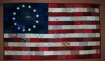
I thought a nice starting place, in light of the election season would be this political quilt. The Donkey quilt was pieced by Beatrice Ruth Calhoun Williamson, Texarkana, in 1932-1934. Some history of donkey quilts can be found here.

Next there is this brightly colored American Tree quilt, circa 1940, made by Herma Williams.

This Pine Cone quilt, pieced by Oscar Evans, circa 1984, is very heavy! I recall that from sewing on the "sleeve". It is much more impressive in person than in this picture.

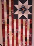
I really like this one (well, I like them all). This Stars quilt, circa 1890-1910, was made by Asia Cummings Shed. It was considered a “show piece,” used only on special occasions.

I hope you will go to the Old State House's web site and look through their on-line exhibit--A Piece of My Soul, Quilts by Black Arkansans. It's very interesting. You could easily spend an hour or two there. I know I'll be back to the site more than twice!

Friday, January 18, 2008

Eye Exam in Facial Trauma

 Updated 3/2017 -- photos and all links removed as many no longer active and it was easier than checking each one.

I perhaps should have done this sooner in the facial fracture series, but am doing it here before doing the orbital fracture post.
In major facial trauma, 15-20% of patients suffer vision-threatening injuries. Early identification and management of such ophthalmic insults often improve the visual prognosis. Manipulations during facial fracture repair can exacerbate unrecognized eye trauma. In addition, ophthalmic problems not documented before facial reconstruction may be interpreted as direct complications of surgery. For these reasons, all physicians who treat patients with trauma above the mandible should appreciate the fundamentals of ophthalmic evaluation and emergency management. This post is to educate myself and others, but it should not be viewed as a substitute for consultation by a qualified eye care provider when an ocular injury is strongly suspected. Eye exams are often not easy in trauma patients, so be sure to document what you are able to do and see, as well as what you are unable to do. A good example is the unconscious patient who will not be able to participate in the visual acuity portion of the exam.

THE EYE EXAMINATION

VISUAL ACUITY
In the United States, normal visual acuity is designated as 20/20. This indicates that a person being tested can see at 20 feet away what the average person can see at this same distance. In Europe, where the testing distance is 6 m, normal vision is depicted as 6/6.
Decreased visual acuity of 20/50 indicates that an individual being tested can see at 20 feet what an average person can see at 50 feet, whereas someone with spectacular 20/10 vision can see at 20 feet what the average person must be 10 feet away to see.
In youth, the intraocular lens is able to change shape and thicken, increasing refractive power and allowing the eye to focus on close objects in a complex process called accommodation.

The ability to accommodate starts to diminish (presbyopia) in most people older than 40 years; thus, there is the need for extra convergence lens power, or reading glasses. As a direct result, the majority of people older than 40 years who are screened in emergency departments using a near-vision card without corrective reading lenses will test as having diminished vision, even though their distance vision and eyes may be perfectly normal.
In addition, consider the relative apparent size of a thread held at 6 inches from the eye versus 3 feet away. Realize that a near-vision card is standardized for use for a standard distance (ie 33 cm or 40 cm) from a subject's eyes. If the card is held closer, the numbers will appear larger than they should be. Likewise, if a presbyopic individual holds the card farther than precisely 33 cm to bring the numbers into better focus, the characters will be effectively smaller and decreased vision may be recorded.
Visual acuity is perhaps the best single test of overall ocular integrity and function, accurate testing is very difficult (if not impossible) in the emergency room setting. Eyelid or orbital swelling may minutely deform the shape of the eye, temporarily altering a patient's refractive need and leading to spuriously low vision testing, even in the presence of usual corrective lenses. Serious ocular injuries, such as a penetrating scleral laceration or a peripheral retinal tear, may have no immediate effect on central visual acuity. Bad vision in the emergency room often results from simple things such as lack of appropriate spectacle correction, blood or mucus in the tear film, and poor effort resulting from anxiety, pain, or intoxication. An eye that can see no light at all, however, is a clear indication of severe ophthalmic injury, unless, of course, the patient is malingering (a complicated topic left to others).
EXTERNAL EXAMINATION
Gross inspection of the eye and ocular adnexa should be part of every examination. Lacerations and contusions over the lateral eyebrow or in the mid glabella are worrisome for the association with posttraumatic optic neuropathy (discussed below). Eyelid lacerations may be full-thickness with underlying globe injury. Remember that forced closure of the eye, such as might occur in anticipation of a blow, creates upward rotation of the globe behind the closed eyelids, known as Bell's phenomenon, in about 75% of the normal population. So when exploring a through-and-through upper eyelid injury, one must not forget to examine the inferior corneal limbus, often hidden beneath the margin of the lower eyelid. Fat prolapsing through an eyelid laceration is strongly suggestive of orbital penetration. Foreign bodies may be difficult to identify in the fat tissue.

Blunt trauma to the eyelids may result in a medial, full-thickness eyelid tear. The medial canthal tendon is a relative, focal weak are and often such an injury will lacerate the canaliculus. Posttraumatic telecanthus usually indicates a nasal side wall fracture with the bone fragment and the still attached medial canthal tendon insertion moving laterally together.
OPTIC NERVE FUNCTION
In the verbal, cooperative patient, one of the best tests of overall optic nerve function is subjective red color saturation. A bright red object is presented to one eye at a time. The patient is asked whether the object's color is of equal hue and intensity in each eye. If an optic nerve has suffered significant injury, ipsilateral color perception will be altered. The red object may appear more dull, orange, or brown than it does with the contralateral eye. Even patients with mild color appreciation deficiencies (8 percent of male subjects in the United States) will be able to tell a difference.
A similar white light intensity test is performed by shining a bright white light in one eye and then the other. Although this is less sensitive than the red color test, it is more useful if there has been trauma within the eye. An eye filled with blood will see approximately the same light intensity as an eye that is not. Also, eyes with unequal-sized pupils may appreciate differences in the low ambient illumination of a red color test, whereas the brighter stimulus of a white light may be seen erroneously as even brighter in an eye where the pupil cannot constrict, casting doubt on the ocular function in the contralateral eye. Importantly, both red and white light saturation tests may yield false-normal results if there is equal compromise of both optic nerves.
PUPIL EVALUATION
In the nonverbal or uncooperative patient the pupil examination may be the only available measure of total ocular function. There are three parts to a pupil examination.
  • First, the size and shape of each pupil should be recorded. An irregularly shaped pupil (corectopia), especially a teardrop- shaped pupil, should raise concern about an anterior penetrating injury to the eye. The point of the teardrop may point toward a laceration hidden beneath conjunctival swelling (chemosis), where the iris has become incarcerated and sealed the wound.
  • The second part of the pupil examination is an assessment of the reactivity to bright light. Each pupil should be viewed independently. A grading system understood by all non- ophthalmologists is trace, sluggish, and brisk. Then, one should swing the light from one eye to the other and back to determine whether there is a relative afferent pupillary defect. An easy way to test this is to watch just one pupil, for example, the left. One should shine the light in the left eye and watch the degree of constriction. While still watching the left pupil, one should move the light into the right eye. The left pupil should minimally dilate in the time it takes to move the light from the left eye to the right, but should then constrict to the same degree as when the light was shined directly into the left eye. If the left pupil does not constrict as well when the light is shined in the right, then there is a problem somewhere along the right visual pathway (retina, optic nerve, optic chiasm, or optic tract). Very nice explanation of anisocoria and abnormal light reflexes by Dr Jeff Mann here.

  • The third part of the pupil examination is assessment of miosis during near synkinesis, often erroneously referred to as pupillary accommodation. This test, in fact, is of little value when evaluating eye trauma, especially if the remainder of the pupil examination is normal, and more properly belongs as part of a complex neurologic evaluation.
Pupil size has no relationship to optic nerve function or visual potential. Instead, pupil size is determined by sympathetic fibers traveling along cranial nerve V and parasympathetic fibers traveling along the inferior division of cranial nerve III, whereas vision is dependent on cranial nerve II, the optic nerve. A totally blind eye is likely to have a normal sized pupil, and an eye with a blown, dilated pupil may have totally normal vision. Also remember that many medications seen in the emergency room setting, such as narcotics and recreational drugs, affect pupil size.
In the setting of trauma, carefully performing and recording a pupil examination is always critical. Too often, PERRLA (pupils equal, round, react to light, and accommodation) is jotted on the chart. A better description might be pupils: 4 mm, round, briskly, and equally reactive to light.
VISUAL FIELD DETERMINATION
In the emergency room, visual field testing in an awake and fully cooperative patient can be more revealing than measuring visual acuity. There are three parts to the visual field assessment.
  • The first, central visual field, evaluates overall central retinal (or macular) function. There are several formal testing mechanisms, such as the Amsler grid. A simplified test is to stand roughly 2 feet from the patient, cover one of the patient's eyes, ask the patient to focus on your nose, and while doing so, the patient should be able to see all the features of your face, including your ears, without any dark or blurry spots. Repeat this test for the patient's second eye.
  • Secondly, to assess peripheral visual field, position yourself 2 to 3 feet in front of the patient with both of you covering mirror eyes (if the patient covers the left eye, you should cover your right eye). Then, with your contralateral hand equidistant between your two heads, bring a wiggling finger in from the far periphery. Have the patient say when the moving finger is first visible. You and the patient should see the finger at approximately the same time. This assumes that you don't have any peripheral vision problems.
  • The third part of visual field analysis, double simultaneous confrontation, more properly belongs as part of a complex neurologic evaluation.
PENLIGHT EXAMINATION
With a penlight, an assessment should be made of the conjunctiva, the cornea, the anterior chamber, and the lens. The conjunctiva, the thin mucous membrane covering the eye, runs from the edge of the cornea, across the surface of the eye, and up the insides of the upper and lower eyelids almost to the eyelashes. Vascular engorgement in the conjunctiva gives a red eye or pink eye appearance that is nonspecific for ocular surface irritation. The conjunctival examination should focus on identifying any foreign bodies, tears in the conjunctiva, and chemosis (conjunctival swelling, either pale or hemorrhagic). Although most subconjunctival blood results from a simple bruise on the surface of the eye, focal globe penetration below the blood must be ruled out. Pale (nonhemorrhagic) chemosis may also represent an occult globe rupture with subconjunctival accumulation of ocular aqueous from the anterior chamber.
The cornea and lens should appear clear, with intact red reflex through both. Clouding of the cornea most likely suggests either old scar or acute infection, and clouding of the lens is generally a cataract. Some types of posttraumatic cataracts can develop acutely.
The anterior chamber, the area between the cornea and the iris or lens, should likewise be totally clear, and there should be an appreciable depth to the front part of the eye. If the anterior chamber is flat, with no space between the cornea and the iris, an occult globe rupture must be suspected. Red or white blood cells filling the anterior chamber are called a hyphema or hypopyon,  respectively.

INTEROCULAR PRESSURE MEASUREMENT
Most techniques for evaluating intraocular pressure are not amenable to being performed by an infrequent examiner. Very high intraocular pressure induced by orbital hemorrhage should not be managed by passing a needle into the anterior chamber. This maneuver lowers intraocular pressure only temporarily and then results in a flat anterior chamber, which may occlude the trabecular meshwork, block aqueous outflow, and create even higher intraocular pressure. If an ocular laceration is suspected, call the ophthalmic specialist.
MOTILITY
The first part of a motility examination determines whether both eyes work together while the patient is looking straight ahead in primary gaze position.
  • If there is sufficient vision to see a finger or pencil held at 3 feet, and the vision is roughly equal in both eyes. It is enough to simply ask a cooperative patient whether or not he or she sees one image. The finger should be held first vertically and then horizontally to check for diplopia in both primary meridians. If diplopia is present, then record the approximate test distance and type of diplopia (e.g., vertical, horizontal, torsional, or a combined).
The second part of the motility examination tests eye movement in each of six major gaze positions: left, right, up and in, up and out, down and out, and down and in.
  • Most significant movement disorders are identified by simply testing up, down, left, and right gaze. Clearly record which movement are normal or abnormal in the medical record. For example, if only four positions of movement are tested, a plus sign should be noted, as opposed to an H, if six positions are examined. For the uninitiated, simply writing the left eye has trouble in up-gaze is probably adequate.
  • When evaluating orbital trauma, a carefully performed and documented ocular motility assessment is critical. Whereas globally restricted movement may indicate diffuse orbital swelling needing delay before surgery, movement more limited in one meridian (e.g., up-gaze and down-gaze) is worrisome for an inferior rectus entrapment that may require urgent orbital exploration and fracture repair to avoid muscle ischemia and permanent dysfunction.
  • Children younger than 12 years, and certainly those younger than 6 years, are at risk for developing amblyopia if they have prolonged dysmotility and do not use their eyes together.
FUNDUS EXAMINATION
Examination of the retina, optic nerve, and retinal vessels is the most technically difficult part of an eye evaluation, especially through an undilated pupil with a direct ophthalmoscope. Dilating the pupil with 1% Mydriacyl (Alcon, Hünenberg, Switzerland) and 2.5% phenylephrine can greatly facilitate the examination.
NOTE: If there is any question about vision, optic nerve injury, visual field, or general neurologic status, dilating drops should not be placed in both eyes, so that pupil function can be assessed independently by other physicians. However, the presumed pathologic eye can usually be dilated without problems, but one should be certain to note in the chart and advise nursing personnel that one pupil has been dilated.
If an accomplished observer finds that there is a good red reflex but no view of the retina, perhaps the posterior chamber is filled with blood. A large white or pale area in an otherwise red retina may indicate retinal ischemia or ocular contusion. A retinal detachment may appear as large undulating, pale folds. Likewise, finding that different parts of the retina are out of focus as compared with others while the power correction in the direct ophthalmoscope is held constant suggests the retina is sitting at different levels. Optic nerve avulsion (picture below) is a rare clinical entity that occurs when there is partial or complete tearing of the optic nerve from the globe at the level of the lamina cribrosa.

IMAGING

The single most useful imaging study in the setting of trauma is probably computed tomography. Relative to computed tomography, plain films are often not as sensitive. Magnetic resonance imaging is contraindicated in the presence of possible metal foreign bodies, more expensive, often more difficult to obtain quickly, and does not show bone as well.
For the orbit, 3-mm computed tomographic sections are usually sufficient, unless a small foreign body is sought. Coronal images are preferred over axial images, although the combination is most helpful.

TRUE OPHTHALMIC EMERGENCIES

Although there are many ocular insults that require rapid attention, the two ophthalmic emergencies where every minute may count are chemical exposure (particularly alkali burns) and ophthalmic infarct.
Industrial chemicals are more likely to be acids, whereas household solutions are more likely alkaline, and the latter are generally more dangerous for the eye. The treatment in either case is
  • copious irrigation with any pH-neutral solution such as water, half-normal saline, or even lactated Ringer's solution. In the presence of severe chemical burns, 10 to 20 liters of irrigant may be appropriate. One way to determine when enough irrigation has been performed is to check for a pH of 7 in the inferior ocular fornix, wait 10 minutes, and check again. In the absence of narrow-range pH paper, a urine dipstick (trimmed if necessary) may offer some indication.
  • Placing a topical anesthetic in the eye first will greatly facilitate the process.
  • Irrigating contact lenses should be used with caution, as they can trap injurious chemical particles.
  • Remember, a bone-white appearance to the conjunctiva may be a bad prognostic sign, indicating severe ocular surface ischemia.
Based on studies in monkeys, the best hope for vision return after a stroke to the eye comes with intervention implemented within 1.5 hours of the insult. In an ophthalmic infarct, the patient reports sudden vision loss, and the only objective finding is an afferent pupillary defect. Other insults, such as posttraumatic optic neuropathy, may present in this fashion, but a vascular accident must always be considered. Intervention should be individualized and directed by someone trained in managing this acute emergency (call the ophthalmologist).

ORBITAL COMPARTMENT SYNDROME

Any sudden increase in orbital pressure can create a compartment syndrome leading to arterial compression or spasm and ophthalmic stroke. In the presence of an orbital fracture with sinus communication, air from nose blowing or sneezing may be forced into the orbit. When the pressure drops after the sneeze, orbital fat falls back into the bone defect, acting as a ball valve and trapping air. A sudden increase in air volume in the enclosed orbit results in sudden increased orbital pressure. Treatment in vision-compromised patients consists of expeditious air evacuation, either through open surgical technique or computed tomography-guided needle aspiration. Alternatively, lysis of the lateral canthal tendon (cantholysis) can rapidly decrease orbital pressure. This requires very little skin incision and fully releases the eyelid when performed correctly.
Retrobulbar hemorrhage (picture below) occurs when an orbital vessel ruptures and leaks blood products into the orbit. This is another form of a compartment syndrome as the orbital space is a closed environment. Any added contents will inevitable increase the pressure inside the orbit and have the potential to negatively impact the ocular structures.

In the setting of an active orbital hemorrhage, however, cantholysis must be performed with caution, as continued bleeding with growing posterior pressure may lead to progressive proptosis and stretch optic neuropathy (or even very rarely partial optic nerve avulsion). The most likely culprit of such heavy bleeding is the infraorbital artery, although the anterior and/or posterior ethmoidal arteries may also be culpable. In suspected, heavy orbital bleeding, the treatment is emergent orbital exploration to obtain artery control.

POSTTRAUMATIC OPTIC NEUROPATHY

Posttraumatic optic neuropathy is vision loss of any degree from optic nerve injury following head trauma. Causes of posttraumatic optic neuropathy include direct optic nerve injury from penetrating objects, fractured bone fragments impinging on the nerve, nerve ischemia, and nerve compression from intrinsic or extrinsic hematoma or edema. Often, more than one mechanism is involved. Posttraumatic optic neuropathy may affect any or all of the following: visual acuity, visual field, pupillary response, and color perception. Dr.Soparkar feels that red color desaturation is perhaps the most sensitive indicator of posttraumatic optic neuropathy in the emergency room. The management of posttraumatic optic neuropathy remains highly controversial and is beyond the scope of this post. Call the ophthalmologist.

CORNEAL EXPOSURE

Many head trauma patients suffer from multiple medical problems and are unable to protect their eyes because of cranial nerve VII injuries, gross exophthalmos, periocular lacerations, or tissue loss. A suture tarsorrhaphy can be temporarily curative, but the eye becomes hidden, and any complications will go unnoticed. Instead, the frequent use of thick, lubricating ointment is preferred. Pure petroleum jelly (Vaseline) is an inexpensive option. Alternatively, a clear adhesive dressing, as is used to dress intravenous line sites, can be applied directly over the eye. It will not stick to the wet eye but adheres strongly to the surrounding skin and forms an effective moisture chamber.
REFERENCES
The Eye Examination in Facial Trauma for the Plastic Surgeon; Plastic & Reconstructive Surgery. Craniofacial Trauma. 120(7) Supplement 2:49S-56S, December 2007; Soparkar, Charles N. S. M.D., Ph.D.; Patrinely, James R. M.D.
Head, Face, and Neck Trauma: Comprehensive Management By Michael G. Stewart; Google Book
Assessment and Management of Ocular Trauma by Sudeep Pramanik, M.B.A., M.D.; University of Iowa Health Care
Ocular Trauma Management for the Primary Care Provider by Joseph M. Rappon, O.D., M.S., F.A.A.O.

Thursday, January 17, 2008

Le Fort Fractures

 Updated 3/2017 -- photos and all links removed as many no longer active and it was easier than checking each one.

The maxilla is the central keystone of the face. It links the cranial base superiorly with the occlusal plane of the lower jaw inferiorly. The maxilla has 4 processes: zygomatic, frontal, palatine, and alveolar. The maxillary sinus is housed within the maxilla and varies in size depending on the degree of pneumatization.
The fractures can be of significant functional and aesthetic importance. Functional problems can lead to disorders of occlusion, nasal obstruction, and trigeminal-nerve sensation. Aesthetic losses include decreased midface height, facial width, facial projection, and malar eminence. These losses can lead to a dish-face deformity.
Renee LeFort (1901) provided the earliest classification system of maxillary fractures. His model described "great lines of weakness in the face" using low-velocity impact forces directed against cadaver skulls. A discussion of fractures of the maxilla would not be complete without a description of LeFort's work.

LeFort Classification of Midfacial Fxs/Signs/Symptoms
Lefort I or transverse fracture of the maxilla (A in picture)
  • The result is a "floating palate" with mobility of tooth bearing segment of upper jaw
  • Disturbed occlusion
  • Palpable crepitation in upper buccal sulcus
  • ‘cracked pot’ percussion note from upper teeth
LeFort II or pyramidal fracture (B in picture)
  • produces a separation and mobility of the midface
  • Gagging on posterior teeth
  • Anterior open bite
  • Periorbital ecchymosis/hematoma
  • There may be diplopia and /or subconjunctival hemorrhage
  • There may be Infra-orbital nerve damage
LeFort III or craniofacial dysjunction (C in picture)
  • Mobile middle third of face
  • Similar symptoms as LeFort II
  • There may be CSF Rhinorrhea (25-50% of II and III fractures)
Maxillary fractures today are often the result of motor vehicle accidents. These high-velocity injuries many times produce fracture patterns not classified by the standard LeFort system, but are described simply by the anatomic structure fractured and the degree of comminution present.
Ocular injury is also commonly associated with midfacial trauma. In a report by Al-Qurainy in 1991, 90.1% of their 363 patients with midfacial trauma had some form of ocular injury. Of those, 63% had transient or minor ocular injury, 16% moderately severe injury, 12% severe ocular injury (angle recession, retinal or vitreous injury, optic nerve damage). In those patients with ocular injury 2.5% lost vision in the affected eye. This data stresses the need for a high index of suspicion for ocular injuries in patients with midfacial trauma. So a thorough eye exam should be performed including visual acuity, inspection of the anterior chamber and the retina, pupillary reflexes, and extraoccular movements. An ophthalmologic consultation may be indicated.

IMAGING
Plain film x-rays have largely been replaced by CT scans. As with zygomatic fractures, a full facial analysis from the top of the head through the mandible with 1.5 mm axial cuts will allow coronal reformatting without additional scanning. Sagittal reformatting is useful in assessing the effect of a complex orbital fracture on the inferomedial bulge of the orbital floor. If the patient presents with neurologic compromise (head injury or intoxication) and cannot comply with a complete physical examination, it is prudent to obtain a complete scan at the time that the head CT scan is obtained.
MANAGEMENT OF THE AIRWAY
Surgical management of Le Fort fractures will require correction of the occlusion and intraoperative intermaxillary fixation. Nasal intubation and tracheotomy are often the preferred approaches, as they will not interfere with the maxillomandibular fixation. The following table was adapted from a flow sheet found in the Stewart text (5th reference):
DentitionFracture Type
Nasal Airway*Le Fort I Fx
*Le Fort I Fx with Mandible Fx
Oral Airway through portal cut in Cunning splints or dentureEdentulousLe Fort I / nasal Injury
Le Fort II Fx
Le Fort III Fx
Panfacial Fx


Oral Airway with tube displace through spacePartially Dentate with SpaceLe Fort I / nasal Injury
Le Fort II Fx
Le Fort III Fx
Panfacial Fx


Guided Nasal Intubation
*Fix Maxilla / Mandible
*Switch to Oral Airway for Nasal/NOE reduction

Fully DentateLe Fort I/ nasal Injury
Le Fort II Fx
Le Fort III Fx
Panfacial Fx


Surgical AirwayLe Fort I /nasal Injury
Le Fort II Fx
Le Fort III Fx
Panfacial Fx


SURGICAL TREATMENT
The goals of surgical treatment 1)re-establish midfacial height and projection, 2) establish occlusal relationship, and 3) maintain integrity of nose and orbits. All patients with midface fractures are given antibiotics, because these fractures are considered open or compound. Violation of the paranasal sinus or alveolus and open soft-tissue wounds are inevitable sequelae of midface fractures. Antibiotics have been shown to decrease the incidence of infection after midface fractures.
Exposure is crucial in repair of the midface fracture. Generally speaking, a Le Fort I fracture is approached from a sublabial exposure, a Le Fort II fracture is approached with a combination of sublabial and periorbital exposure, and a Le Fort III fracture requires a combination of sublabial and bicoronal fracture for adequate exposure.
The surgical approaches to fractures of the midface have changed radically in the past 20 years. The technology has now evolved to allow for miniplate fixation to the midface instead of bulky external hardware. Complex internal wiring used to be the standard of care 10 years ago, but due to poor cosmetic results and extended periods of IMF, newer technologies (mini-plate technology) have replaced it. If large bone deficiencies are present, bone grafting may be necessary.

The following general guidelines are meant for edentulous patients, but are good start for planning for all. Those with teeth can also be treated with arch bars, etc while their general health improves. Remember most of these patients with have serious (head, neck, chest, etc) injuries also.
No Displacement
  • Soft Diet for 2-3 weeks
  • Incorporation of dentures
Displacement, Patient Stable, Good General Condition
  • Open reduction
  • Reconstruction of midfacial buttresses
  • Miniplate osteosynthesis with simultaneous preprosthetic surgery if necessary
Displacement, Patient in poor general condition
Le Fort I Level--
  • No active therapy
  • Le Fort I osteotomy later
Le Fort II or III Level--
  • Minimal therapy, repositioning of nose and orbital rim
  • Le Fort I osteotomy later

COMPLICATIONS
Early
  • Extensive hemorrhage
  • Airway obstruction
  • Infection--When given appropriate antibiotics, the risk of meningitis is small (less than 1%). Sinusitis occurs in 1-2%.
  • CSF leak
  • Ocular Problems -- Approximately 17-25% of patients with severe fractures will suffer some type of ocular problem--blindness (1-2%), diplopia, enophthalmos, epiphora, blurred vision, lacrimal drainage problems.
Late
  • Palpable or exposed hardware
  • Non-union/ Malunion
  • CN V2 Anesthesia -- The incidence of an immediate postoperative infraorbital nerve deficit from Le Fort I and II fractures has been reported at 65%. It is directly related to fracture displacement of 1 mm or more. Patients with less than 1 mm displacement regain normal sensation after 3 months. 30% of those with displacement of greater than 1 mm have a persistent sensory deficit at 1 year.
  • Disturbances of Smell and Taste may occur in 23-38% of patients with up to 80% of Le Fort III patients reporting disturbance of smell.
  • Extra-Occular Muscle Imbalance
  • Malocclusion may result from fixation of improperly reduced fractures or insufficient fixation.
  • Secondary Facial Deformities result from inadequate reduction and stabilization of the nasal bones. Nasal deformities are reported in 12-33% of upper midface fractures.
REFERENCES
Le Fort Fractures by Anil R Shah, MD and Galdino Valvassori, MD --eMedicine Article, Oct 27, 2006
Facial Trauma, Maxillary and Le Fort Fractures by David W Kim, MD and Kristin K Egan, MD --eMedicine Article, June 8, 2006
Maxillary and Periorbital Fractures by Gordon Shields MD and Francis B Quinn MD; Grand Rounds Presentation, UTMB; January 7, 2004
Atlas of Emergency Medicine; Kevin J. Knoop, Lawrence B. Stack, Alan B. Storrow; Google Books
Head, Face, and Neck Trauma: Comprehensive Management; Michael G. Stewart, MD; Google Books
Facial Fractures; Core Curriculum Syllabus: Emergencies in Otolaryngology-Head and Neck Surgery; Baylor College of Medicine
Facial Fractures: The Upper Face by Dough Humphreys, MD; Dalhousie University (PDF file)
Maxillo-facial Injuries; PatientPlus

Wednesday, January 16, 2008

What are the odds?

It was a quiet late summer Saturday afternoon. The pager went off. The message--Call TT at 555-5555.
I always do a mental check list of possible problems when I recognize the patient's name. This one had had a tummy tuck done 6 weeks previously. She had healed with no problems or issues. She hadn't asked for a refill on pain medication. All of her follow up visits had been routine. I couldn't begin to image why she was having me paged.
"TT, this is Dr. Bates. How can I help you?"
"Dr. Bates, my incision has popped open!"
"Slow down and take a deep breath. Try to stop crying and repeat what you just said. I'm not sure I understood you."
"I fell when I stepped out of the boat. I landed on some gravel and my incision on my right side has opened up."
"Okay, where are you and how long do you think it would take to get to my office? The building is locked because it's Saturday, but I can get us in. I'll look at you there and if I can I'll fix it for you there, I will." No need to further stress her with the possibility of surgery at the hospital. She was already thinking of the added cost of this, having maxed out her savings and credit to have the initial surgery. "Just come on"
"It'll take us about two hours. I'm all dirty. I need to change clothes."
"Don't worry about your clothes, just come."
Boy, was I surprised! As my husband likes to say, "you can't make this stuff up". This is not a "complication" that you warn patients about after surgery. This is different from wound dehiscence. This literally was a healed wound that had incurred the pressure of the fall onto one rock with the point of maximum force right over the healed scar at her right hip. Scars at 6 weeks are only at approximately 55% of the final strength that will be reached at approximately 10-12 weeks post-injury. Even then scars only have 80% of the tensile strength of uninjured skin. But what are the odds.........
When they arrived at my office, I found an open wound about 18 cm long, gaping nearly 5 cm at the widest point. It was centered over the right anterior hip bone. There was dirt, grass, and small gravel in the wound. There was no active bleeding . I got her to lay down on the exam table and did a local block using 0.5% Lidocaine with Epi and 0.5% Marcaine without Epi. Then I thoroughly cleaned the wound out with normal saline and Betadine solution, picking out the grass and gravel. Cleaned some more. Then I closed the wound with vicryl and PDS. I reassured her that most likely in two years, we wouldn't be able to tell which part of the scar had been reopened. I sent her home on antibiotics, but she declined pain medicine (had some left over from the surgery). Both TT and I were relieved that it wasn't any more serious than it was.
Now when patients ask for 100% guarantees of their postop courses, I use this example of how even best laid plans can be changed by "life", the "universe", whatever. Some things are not in our control. I tell them I will do everything I can to ensure a good outcome. I ask them to help me by following instructions and using common sense. But what are the odds.....

Tuesday, January 15, 2008

Mandible Fractures

Updated 3/2017 -- photos and all links removed as many no longer active and it was easier than checking each one. 

The primary causes of mandible fractures, like other facial fractures, are vehicular accidents and assaults. Other significant causes are falls and sports injuries. In the US, the mandible is the third most fractured bone of the face. There is an adult male to female ration of 3:1. It is reduced to a ratio of 3:2 in children.
Incidence of other major injuries is as high as 50% in high-impact mandibular fractures, whereas it is 21% in low-impact fractures. This includes an associated cervical spine injury rate of 0.2-6%. Mortality rate in high-impact fractures is as high as 12%, yet death rarely results directly from maxillofacial injury.
Location of fractures
Different mechanisms are associated with varying locations. Fractures from automobile crashes most frequently occur at the condyle and symphysis, those from motorcycle accidents at the symphysis and alveolus, and those from altercations mostly at the condyles, angles, and body.
Most fractures occur in the body (21-29%), condyle (26-36%), and angle (20-25%) of the mandible. The parasympheseal and symphyseal fractures account for 15-17% of mandibular fractures, whereas the ramus (3-4%) and coronoid process (1-2%) have a lower occurrence rate.

Mandibular fractures are isolated (no other facial fractures) in an average of 70% of the patients. Of the patients reported, 15% had another facial bone fracture along with the fractured mandible. A mean of 53% of patients had unilateral fractures, 37% of the patients had 2 fractures, and 9% had 3 fractures.
Mandibular fractures usually occur in 2 or more locations because of the bone's U shape and articulations at the temporomandibular joints (TMJ). Fractures also may occur at a site apart from the site of direct trauma. A large percentage of mandibular fractures are open, as they often fracture between teeth and communicate with the oral cavity.
DIAGNOSIS
Examination of the patient with a mandibular fracture should include:
  • Assessment of the occlusion
  • Palpation of the mandibular contours
  • Bimanual manipulation of the occlusal segments to detect fracture mobility
  • Voluntary mandibular mobility, including maximal opening and excursive movements. Deviations on opening should be recorded.
  • Examination of dentition for injuries/avulsion of the teeth. Make sure each tooth is accounted for.
  • Soft tissues adjacent to the fracture should be examined for hematoma, lacerations, and the integrity of the attached gingiva.
  • Note any neurosensory disturbance in the mental nerve (lower lip and chin) distribution. Fractures that cross the inferior alveolar canal are very likely to result in a change here.
IMAGINING

It is important in imaging of mandible fractures to obtain images that allow evaluation in at least two planes. The following types of radiographs are helpful in diagnosis of mandibular fractures:
    • Panoramic radiograph--shows the entire mandible including the condyles. This combined with a posteroanterior (PA) x-ray is the most commonly used for mandible fractures. Panoramic x-rays are not available in some hospitals.

    • Mandibular series (when panoramic not available)--usually includes
  1. Posteroanterior (PA) mandibular view
  2. Reverse Townes View--This is the plain film of choice for excluding condylar and subcondylar fractures.
  3. Bilateral Oblique Views
  4. Lateral Views
  5. Submentovertex View
  • Temporomandibular joint views including tomography
  • CT scan -- both axial and coronal images should be obtained if possible, if CT scans are deemed necessary.
  • Occlusal views are helpful for accurate assessment of symphyseal fractures.
  • Obtain periapical radiographs of the teeth on either side of the fracture to assess root fractures
CLASSIFICATION
There are many different classifications of mandibular fractures. They are done by type and location of the fracture. There are good reviews of all the classifications here and here.
Closed or Open
  • Is there a fracture that communicated with the extraoral environment through a tear in the mucosa? Does the fracture run into a tooth socket?
Displaced or Nondisplaced
  • The fracture may be displaced as a direct result of the trauma or may be secondary to muscle contraction with movement of the fracture. Most common sites of displaced fractures are the body, symphysis, and angle.
  • Nondisplaced fractures are often seen in the condyle, coronoid process, and ramus. The large muscle masses in these areas serve to stabilize the fractures.
Complete or Incomplete
  • Greenstick fractures are those in which one side of the bone is broken and the other side is bent. These are most common in children.
  • Complete fractures pass entirely through both cortices of the bone. Often the periosteum will remain intact when the fracture is incomplete.
Linear or Comminuted
  • Simple fractures are linear and generally produce only two bone fragments.
  • Comminuted fractures involve many small fragments that are difficult to reduce and stabilize. The vascularity of the fractured segments may be compromised and can lead to nonunions.

TREATMENT
NONSURGICAL
Conservative treatment is indicated when mandibular fracture is nondisplaced and immobile. The occlusion must be normal. The patient may be placed on a soft diet and mandibular function restricted until healing is complete.
SURGICAL
Mandibular fractures should be reduced as soon as possible to minimize pain and reduce the risk of infection.
This table of SUGGESTED MANAGEMENT of Mandibular Fxs is from the Selected Readings in Plastic Surgery, 1994,
Location of FxDisplacementReduction/ Fixation
CondyleMinimal
Moderate
Severe
Closed
Closed
Open, plating or wiring
RamusMinimal
Moderate
Severe
Closed
Closed
Open, plating or wiring
AngleMinimal
Moderate
Severe
Closed
Open, wiring and plating
BodyMinimal
Moderate

Severe
Closed
Open, plating or lag screw (combo)
Open, plating or lag screw
SymphysisMinimal
Moderate

Severe
Closed
Open, lag screw or plating


Some caveats for closed reduction
  • Nondisplaced favorable fractures: Open reduction carries an increased risk of morbidity, thus use the simplest method to reduce and fixate the fracture.
  • Grossly comminuted fractures: Generally, these are best treated by closed reduction to minimize stripping of the periosteum of small bone fragments.
  • Severely atrophic edentulous mandibles: These have little cancellous bone remaining and minimal osteogenic potential for fracture healing. Closed reduction with the use of circummandibular wires offers a more conservative approach.
  • Fractures in children involving the developing dentition: Such fractures are difficult to manage by open reduction because of the possibility of damage to the tooth buds or partially erupted teeth. A special concern in children is trauma to the mandibular condyle. The condyle is the growth center of the mandible, and trauma to this area can retard growth and cause facial asymmetry. Early mobilization (7-10 d of intermaxillary fixation) of the condyle is important. If open reduction is necessary because of severe displacement of the fracture, the use of resorbable fixation or wires along the most inferior border of the mandible may be indicated.
  • Coronoid fractures: These fractures usually require no treatment unless impingement on the zygomatic arch is present.
Some caveats for open reduction
Condylar fractures: Although strong evidence supporting open reduction of condylar fractures is lacking, a specific group of individuals benefit from surgical intervention. Careful evaluation of each case on an individual basis is crucial.
    • Absolute indications
      1. Displacement of the condyle into the middle cranial fossa
      2. Inability to obtain adequate occlusion by closed techniques
      3. Lateral extracapsular dislocation of the condyle
    • Relative indications
      1. Bilateral condylar fractures in an edentulous patient when splints are unavailable or impossible because of severe ridge atrophy
      2. Unilateral or bilateral condylar fractures when splinting is not recommended because of concomitant medical conditions or when physiotherapy is not possible
      3. Bilateral fractures associated with comminuted midfacial fractures

POSTOPERATIVE CARE
The primary concern in patients with mandibular fractures who have been treated with maxillomandibular fixation is the airway. If the cause of the injury was an accident, a nasogastric suction tube is inserted at surgery and left in place for 6 hours to prevent aspiration. It is recommended that the patient not be extubated until fully awake.
It is important for the patient to maintain adequate nutrition. Feedings should progress from a clear-liquid diet to a high-protein, full-liquid diet, to a blended fractured-jaw diet.
COMPLICATIONS
  • Infection
  • Delayed healing and nonunion--most common cause is infection, second most common cause is noncompliance, and then there is inadequate reduction, metabolic &/or nutritional deficiencies.
  • Nerve paresthesia--(Inferior Alveolar nerve) occur in 2%
  • Malocclusion and malunion
  • TMJ problems
REFERENCES
Facial Trauma, Mandibular Fractures by Adel R Tawfilis, DDS and Patrick Byrne, MD-- eMedicine Article, March 10, 2006
Fractures, Mandible by Thomas Widell, MD -eMedicine Article, April 24, 2005
Craniofacial Trauma; Supplement to Plastic & Reconstr Surgery, Vol 120, No 7, Suppl 2, Dec 2007; Larry H Hollier, Jr MD and James F Thornton MD
Facial and Mandibular Fractures, Approaches To Differential Diagnosis In Musculoskeletal Imaging by Michael L. Richardson, M.D.; University of Washing School of Medicine
Mandible Fractures by Karen Stierman, MD and Byron J Bailey, MD --UTMB Grand Rounds, June 14, 2000 (PDF File with nice slides)
CLASSIFICATIONS OF MANDIBULAR FRACTURES-REVIEW; Journal of IMAB - Annual Proceeding (Scientific Papers) 2006, book 2; Hristina Mihailova, Department of Maxillo-facial radiology and oral diagnostic, Faculty of Stomatology, Medical University-Sofia, Bulgaria (PDF file)

Monday, January 14, 2008

Nasoethmoid Orbital Fractures

Updated 3/2017 -- photos and all links removed as many no longer active and it was easier than checking each one. 

Continuing with my review of facial fractures. Today is the area between the eyes--the nasoethmoid orbital region. Nasal fractures represent the third most commonly broken bone in the body, and the nose is the most commonly broken facial bone. There is a nice review of simple, acute nasal fractures here.
A facial fracture is considered a nasoethmoid orbital [or naso-orbital-ethmoid (NOE)] fracture if the fracture involves the bone to which the medial canthal tendon is attached. It is important to distinguish NOE fractures from isolated nasal fractures, orbital rim fractures, and fractures which involve only the ethmoid labyrinth. The medial canthal tendon provides globe (eye) support as part of a suspensory sling, which is in continuity with the lateral canthal tendon, and upper and lower tarsal plates. The tendon is also intimately associated with the lacrimal drainage apparatus.
The naso-orbito-ethmoid complex is composed of a confluence of several bones: (1) frontal bone, (2) nasal bone, (3) maxillary bone, (4) lacrimal bone, (5) ethmoid bone, and (6) sphenoid bone. The key anatomical region is the central bone fragment of the medial orbital rim, into which the medial canthal tendon inserts.
Nasoethmoid fractures typically result from a forceful blow to the central aspect of the midface. Motor vehicle accidents are the most common source of injury, followed by assault. Possibly due to the advent of safety air bags in most newly produced vehicles, the incidence of these injuries is decreasing. Less forceful injuries are needed to cause NOE fractures than zygomatic, maxillary, or frontal fractures.
DIAGNOSIS
The best way to confirm the diagnosis is the combination of physical examination and CT scan. Even with soft tissue swelling, physical examination/observation may be informative.
  • Focal areas of swelling or hematoma may overlie an isolated fracture. Periorbital swelling may indicate LeFort II or III fractures.
  • Palpation may reveal mobile bony segments, step-offs, or crepitus, all of which suggest fracture.
  • If the entire nasal pyramid is posteriorly displaced or "telescoped in", the diagnosis is obvious.
  • Measurements of the intercanthal distance should be compared to the interpalpebral distance of the eyes. If the former is significantly larger than the latter, traumatic telecanthus from NOE fracture is presumed. In patients in whom edema makes localization of the medial palpebral angle imprecise, an alternative measurement is interpupillary distance, which should be approximately double the intercanthal distance. If intercanthal distance exceeds one-half the interpupillary distance, traumatic telecanthus must be considered. Normal values are 60 to 70 mm (interpupillary) and 30 to 35 mm (intercanthal).
  • The eyelid traction test is another test for the integrity of the MCT. To do this test, the examiner grasps the lower lid in question and pulls laterally while palpating the nasal root. A lack or reduction of tension with lateral pull suggests NOE fracture with MCT displacement.
  • A thorough eye examination with visual acuity, pupillary responses, and extraocular motion is crucial. Close inspection of the lower lid may reveal a rounded medial palpebral fissure and lid laxity.
  • Although epiphora (overflow of tears) may be associated with NOE fractures, its presence is an unreliable indicator of injury. Lacrimal function may be assessed more accurately by placing irrigation and probing in conjunction with the Jones dye test of lacrimal function.
  • Any fluid from the nose should raise the possibility of a CSF leak. If enough fluid can be collected, it should be sent for analysis of beta2-transferrin, an indicator for CSF. A cruder test for CSF may be performed by collecting a few drops of fluid on filter paper and examining the pattern of migration of fluid. Blood and water tend to form a central pool, while CSF tends to form a second outer ring.
IMAGING
Patients with suspected facial trauma should initially be evaluated with a complete craniofacial CT scan. A full facial analysis from the top of the head through the mandible with 1.5 mm axial cuts will allow coronal reformatting without additional scanning. Plain radiographs have limited utility in assessing isolated nasoethmoid fractures. These films may demonstrate opacification or clouding of the maxillary and ethmoid sinuses, indicating the presence of blood. They are unlikely to characterize the relatively detailed osseous anatomy of the NOE complex. Therefore, CT scan images have replaced plain films as the main imaging tool to assist in the diagnosis and treatment planning for NOE fractures.

FRACTURE CLASSIFICATION
Markowitz and colleagues classify NOE injuries into three fracture patterns accordingly
Type I--single, noncomminuted, central fragment without medial canthal tendon disruption
Type II--comminution of the central fragment without medial canthal tendon disruption
Type III--severe central fragment comminution with fracture extension through the medial canthal insertion or have avulsed the tendon insertion

SURGICAL TECHNIQUE
A combination of four different incisions may be used to provide complete exposure to these fractures--the coronal, a limited midline, a lower eyelid, and the upper buccal sulcus.
Type I Fractures
  • These can be managed with plate-and-screw fixation. The large single-segment fragment must be reduced and then stabilized at the superior orbital rim and piriform. Sargent avoids the eyelid incision in these.
Type II Fractures
  • These fractures require wider exposure to adequately reduce and stabilize. The most common incisions used for both Type II and III fractures are the coronal, lower eyelid, and upper buccal sulcus.
  • The key bone segment to identify is the medial orbital rim bone, into which the medial canthal tendon (MCT) inserts. Care must be taken not to inadvertently strip off the MCT insertion.
  • The key step in the stabilization of the canthal-bearing bone segments is placement of the transnasal wires to obtain a symmetric, secure reduction of these bone fragments. Sargent states that screw-and-plate fixation is not an effective technique in this particular step of stabilization of either Type II or III. There are some nice pictures of his technique in the PRS supplement, Dec 2007 listed below in the references.
Type III Fractures
  • Sargent states that it is rare for the tendon to be completely avulsed unless there is a penetrating injury over the tendon.
  • If the bony fragment to which the MCT inserts is not large enough to place two drill holes approximately 4 mm apart with several millimeter of surround good bone, it should be replaced with a bone graft so that there is adequate support.
  • If the MCT is partially avulsed and the medial bone fragment is of adequate size, the tendon should be reinforced.
  • A slight over-reduction is often needed and can be easily accomplished by continued twisting of the wires.
Other associated fractures (zygomatic, orbital defects, etc) and will need to be addressed as appropriate.
Commonly, there is telescoping of the nose (bridge of the nose is pushed-in and compressed). This characteristic appearance on profile indicates loss of support and the need for a cantilever nasal bone graft. Simple reduction of the NOE and septal fracture will not be adequate treatment.
COMPLICATIONS
  • Persistent telecanthus postoperatively implies inadequacy of the original technique or detachment of the transnasal wire. Reexploration is indicated in such instances.
  • Injury to the lacrimal system causes obstruction and epiphora. It is best avoided by intraoperative stenting of the lacrimal puncta and duct. Care should be made to avoid securing wires too low on the medial nasal wall and lacrimal crests so they do not impinge on the lacrimal sac.
  • If the transnasal wires pass too low on the septum in repair of a type III fracture, the wires may pull the MCT inferiorly and result in scleral show and lid laxity. Repair requires repositioning of the wires to a higher point on the nasal septum.
  • Lower lid ectropion may follow a subciliary (eyelid incision) approach. If severe ectropion occurs, breaking up the scar with Z-plasty or skin grafting from the opposite lid skin may be necessary.
  • Nerve injury may have occurred prior to surgery from the initial traumatic insult. Therefore, the status of the main sensory and motor nerves of the face and forehead must be documented prior to surgery. Care should be taken to identify and preserve the supraorbital and infraorbital neurovascular pedicles while the soft tissue flaps are raised. More commonly, supraorbital nerve injury results from nerve stretching when retracting the soft tissue and orbital tissues to gain access to the superior and medial orbital rims.
  • The nose may be foreshortened, with lack of projection and contracture of the soft tissue.
REFERENCES
Craniofacial Trauma; Supplement to Plastic & Reconstr Surgery, Vol 120, No 7, Suppl 2, Dec 2007; Larry H Hollier, Jr MD and James F Thornton MD
Facial Trauma, Nasal Fractures by Vipul R Dev MD and others--eMedicine Article, Oct 3, 2006
Facial Trauma, Nasoethmoid Fractures by David W Kim MD and others -- eMedicine Article, May 26, 2006
Nasoorbitoethmoid Fractures by E Bradley Strong MD and others -- eMedicine Article, April 30, 2007
Naso-orbital-ethmoid Fractures by Michael G. Stewart, M.D.
January 7, 1993; Grand Rounds--Baylor College of Medicine
Naso-orbital Ethmoid and Frontal Sinus Fractures by Jim C Grant, MD and Byron J Bailey, MD-- Grand Round Presentation, UTMB; April 29, 1998 (power point presentation)
Eyelid Anatomy by Bhupendra C K Patel, MD and others -- eMedicine Article, April 11, 2006
Management of the Medial Canthal Tendon in Nasoethmoid Orbital Fractures: The Importance of the Central Fragment in Classification and Treatment; Plastic & Reconstr Surg, 87:843, 1991; Markowitz, B L, Manson, P N, Sargent L A, et al

Sunday, January 13, 2008

SurgeXperiences 113 -- call for submissions

 Updated 3/2017 -- all links removed as many no longer active and it was easier than checking each one.

 Terry over at Counting Sheep will be hosting the next edition (113) of SurgeXperiences on Sunday, January 20, 2008.   She wants to try something a little different  and will be calling the edition "Operating After Hours".  Terry would like you to share any of your surgical-related tales that have occurred after "normal operating hours".  Please send all submissions here.   The deadline for submission of  your articles will be Friday, January 18, 2008. ALL are welcome to contribute.
The current edition is up at The Sterile Eye , a very nice blog.

Saturday, January 12, 2008

First QOV's for 2008

Here are two quilt tops and a finished quilt that I am donating to the Quilt of Valor Foundation. I finished the quilt after Thanksgiving, but never submitted a destination request until this past week. The two tops were finished towards the end of the year as well. There was a note on the QOVF website stating that no requests would be answered, due to a holiday break, until after Jan 6, so I just waited until this week to submit those as well.
This top is 51" X 74".
It is a nine-patch made up of left-over green fabrics
The figures are Elvis silhouettes.
This patriotic quilt is 51" X 68".
It is a variation of a square in a square.
Here is a closer view.
This is the Sunshine and Shadow quilt I started
after Thanksgiving. It is 54" X 68".
Here is a closer view.

Friday, January 11, 2008

Zygomatic Fractures

 Updated 3/2017 --photos and all links removed as many no longer active and it was easier than checking each one.

I wish I was good at facial fractures. I am not. There wasn't enough volume of these cases when I was a plastic surgery resident, so I came into private practice not feeling comfortable with them. Then there wasn't much volume in the early years of my practice to gain a comfort level with them. Later I began to avoid them. When EMTALA came along, I decided to withdraw my facial fracture privileges from the hospitals where I continue to do unassigned ER call. I felt this was my only way to protect the patient from my lack of skill or insufficient skill or however you want to put it. I continue to sew up the facial lacerations, but if there is a fracture associated with the laceration the ER now has to call someone else.
Before EMTALA, I would have sewn up the lacerations and gotten the appropriate X-rays (now CT scans). Then the next day, I would have called someone else--either plastic surgeon, ENT, or oral maxillary surgeon to take over the care. They wouldn't have been called in the middle of the night, but during the day time hours. It would have been okay. No one would have felt like I was "dumping" on them.
Still I continue to ready the journal articles about facial fractures. As you can see from the second reference below, there has been a recent supplement volume (very comprehensive) dedicated to these fractures. Since I learn by writing these posts, I thought I would try to tackle this topic over several posts. It still won't improve my OR skill with them, but it might clarify some of my understanding of them. I hope you will indulge me and maybe learn with me.
There is a nice basic summary at eMedicineHealth on facial fractures.
ZYGOMATIC FRACTURES
The zygoma is the bone that is known as the cheek bone. It articulates with the maxilla, the temporal bone, the sphenoid bone and the frontal bone. It forms the prominence of the cheek and part of the lateral wall and floor of the orbit (eye socket). It is this prominence that makes it so susceptible to trauma.
The mechanism of injury usually involves a blow to the side of the face from a fist, object (like a baseball), or secondary to motor vehicle accidents. Studies show that 80% of these injuries are due to motor vehicle accidents. Moderate force may result in minimally or nondisplaced fractures at the suture lines. More severe blows frequently result in inferior, medial, and posterior displacement of the zygoma. Comminuted fractures of the body with separation at the suture lines (the places where the different bones meet/join) are most often the result of high-velocity motor vehicle accidents.
Over the years, isolated fractures of the zygoma have been called zygomatic, tripod, or orbitozygomatic fractures. The last term is the preferred. Here in the United States, zygomatic fractures are the second most common fracture of the facial bones. The first being nasal bone fractures. As many as 5% of these patients have associated ophthalmic injuries. Males are afflicted more commonly than females by a 4:1 ratio. Most cases occur in young patients in their second to third decades of life.
DIAGNOSIS
Physical signs and symptoms of zygoma fractures include
  • Subconjunctival hemorrhage and periorbital ecchymosis are seen in as many as 50% of patients.
  • Disturbance of sensation in the region of the infraorbital nerve
  • Palpable step-offs in the upper lateral orbital rim, inferior orbital rim, and upper buccal sulcus
  • Emphysema within the orbit or overlying soft tissues of the cheek
  • Trismus -- is difficulty with mastication and can occur because of masseter spasm or bony impingement of the coronoid process.
  • Malposition of the globe and /or diplopia (double vision)--Diplopia may be secondary to change in pupillary alignment or to entrapment of the inferior rectus muscle by the orbital wall fracture. Patients with entrapment may complain of diplopia on upward gaze.
  • Globe injury needs to be ruled out as well. Traumatic optic neuropathy is a complication that has been reported in about 2 to 5 percent of severe facial trauma.
Facial swelling is almost always substantial by the time the patient is seen by the surgeon. This tends to minimize the degree of deformity, masking the cheek malposition. The swelling may take 2 weeks to resolve so that the patient (and surgeon) can visually (not by CT scan) appreciate the true deformity. However, the best result is obtained if the reduction and stabilization of the fracture is done within the first 2 weeks after the injury. So the surgeon has to be good at 3-D visualization to fully appreciate the bone structure injury and to optimally reduce the fracture while the soft tissue swelling is still present.
SECONDARY DEFORMITIES
These are deformities that occur because of untreated or mistreated fractures. They are not uncommon (unfortunately). They include
  • Underprojection of the cheek and a wide face.
  • Overprojection is possible, but not as common as the first.
  • Globe malposition--either enophthalmos (a sunken eye, is the most common) or exophthalmos (eye protrudes out of the socket).
  • Soft-tissue deformities from inadequate suspension after extensive surgical exposure. Cheek ptosis, inferior displacement of the lateral canthus, temporal hollowing, and lower lid ectropion are the most common.
IMAGING
Patients with suspected facial trauma should initially be evaluated with a complete craniofacial CT scan. A full facial analysis from the top of the head through the mandible with 1.5 mm axial cuts will allow coronal reformatting without additional scanning. Sagittal reformatting is useful in assessing the effect of a complex orbital fracture on the inferomedial bulge of the orbital floor. If the patient presents with neurologic compromise (head injury or intoxication) and cannot comply with a complete physical examination, it is prudent to obtain a complete scan at the time that the head CT scan is obtained.
CLASSIFICATION
Several classifications of zygomatic fractures have been described in the literature, but none seem to be universally accepted. Most classifications are based on the degree of comminution, whether the fracture is simple or compound, and the site of the fractures. In 75% of cases, these fractures are displaced inferiorly, medially, and posteriorly. The classification system by Knight and North identifies six groups:
  • Undisplaced -- require no therapy, see Medical Care below
  • Arch fractures -- considered a stable fracture
  • Unrotated body fractures -- can usually be treated by closed reduction
  • Medially rotated body fractures -- considered an unstable fracture
  • Laterally rotated body fractures -- considered a stable fracture
  • Complex fractures having additional fracture lines across the zygomatic body
MEDICAL CARE
The literature indicates that 10-50% of all zygomaticomaxillary complex fractures require no surgical intervention. This is suitable for fractures that are nondisplaced or minimally displaced or where systemic status precludes operative intervention.
  • Stable, nondisplaced fractures may be observed weekly for healing.
  • Avoidance of nose blowing is mandatory in the medical care of these patients. The disrupted orbital walls can allow air to be forced into the retrobulbar space and cause pain and visual loss. This avoidance of nose blowing should last for several weeks to a couple of months.
  • Avoid sleeping face down or with pressure on the affected side of the face
  • The routine use of systemic antibiotics for isolated zygomatic arch fractures generally is not recommended.
  • Patients commonly are placed on a soft diet for six weeks. A dietary consultation may be warranted.
  • All contact sports and most strenuous activity also should be avoided for several weeks.

BASIC PRINCIPLES OF SURGICAL REPAIR
The aims of treatment of zygomatic complex fractures include the restoration of normal facial form, normal sensory nerve function, normal globe position, and normal masticatory function. Indications for repair of zygomatic complex fractures include displacement or instability of the fracture, mechanical restriction of mandibular movement (chewing), alteration in facial contour, globe dystopia, enophthalmos, diplopia, or sensory nerve deficit.
Generally, it is suggested to avoid surgery during times of maximum edema but prior to the adhesion of displaced bony fragments and scarring of soft tissues into bony defects. Most surgeons advise surgical intervention prior to the formation of dense scar tissue. As a general guideline, surgery should be undertaken prior to 3 weeks postinjury, optimally prior to 2 weeks postinjury.
Isolated arch fxs and minimally displaced noncomminuted fxs
  • The Gilles approach may be used. It can be done under either local anesthesia with sedation or under general anesthesia. A vertical incision is made in the temporal hairline, and the elevator is then tunneled underneath the temporalis fascia, keeping in mind the superficial temporal artery branches and the temporal division of facial nerve, which is nearby. Then lateral pressure with superior anterior rotation is exerted to pop the fracture back in place.
Unstable or potentially unstable fractures
  • A more aggressive approach using open-reduction techniques and rigid stabilization with plating systems (eg, Synthes or Leibinger) is the standard of care today. This approach provides direct access to the frontozygomatic suture, orbital floor, and infraorbital rims.

  • Precise reconstruction with rigid internal fixation of the zygoma at 2 or 3 points (across the frontozygomatic suture, the inferior orbital rim, and the lateral midfacial buttress) is needed to counter the force of the masseter muscle. The orbital contents can be supported as for simple orbital floor fractures.
COMPLICATIONS of Surgical Repair
Many of these are included in the list of secondary deformities listed above.
  • Diplopia has been quoted to occur between 3.4 and 8 percent. It is most often thought to be secondary to extraocular muscle contusion, swelling, and occasionally entrapment as well. As a result, forced duction test is recommended prior to closure to make sure there is no muscle entrapment, and when in doubt CT can be used.
  • Anesthesia/dysesthesias, usually in the infraorbital nerve region, has been reported between 0.4 and 11.6 percent postoperatively as well; however, usually they resolve on their own, but if they last more than three to twelve months, then one may suspect impingement as a possible cause.
  • Enophthalmos, as you see in this picture here, is most often secondary to inadequate fracture reduction. This has been reported to occur anywhere between 1 and 23 percent postoperatively. If enophthalmos is noted during the early postoperative period, reoperation is a viable option. Most often, the enophthalmos is thought to be due to the fact that the zygoma has been fixed too far posteriorly and laterally; however, in the late postoperative period, reoperation will often require osteotomy (re-fracture).
  • Complications related to hardware eventually requiring removal is also not uncommon. The most commonly cited reason for needing removal is palpability, specifically in the infraorbital rim and the frontal zygomatic suture region. In this study, they also saw an isolated case of plate fracture.
  • The rare complication of sudden onset blindness resulting from retrobulbar hemorrhage following reduction of even simple zygomatic fractures means that in some instances, this procedure may be unsuitable for outpatient surgery. This serious complication, although rare (0.3% of treated zygomatic fractures), is potentially reversible upon early recognition of the symptoms and signs of retrobulbar hemorrhage (eg, pain, proptosis, loss of vision, decreased motility). If the surgeon suspects a retrobulbar hemorrhage, a lateral canthotomy and cantholysis should be performed as soon as possible. This should be completed at the bedside if the patient has visual compromise and is not near the operating suite.
REFERENCES
Facial Trauma, Zygomatic Complex Fractures by Zachary Segal MD, et al--eMedicine Article
Craniofacial Trauma; Supplement to Plastic & Reconstr Surgery, Vol 120, No 7, Suppl 2, Dec 2007; Larry H Hollier, Jr MD and James F Thornton MD
Orbital Fracture, Zygomatic by Stuart R Seiff, MD, et al--eMedicine Article
Facial and Mandibular Fractures by Michael L. Richardson, M.D., University School of Medicine
Blindness after Reduction of Facial Fractures; Plastic & Reconstructive Surgery. 102(6):1821-1834, November 1998; Girotto, John A. M.D.; Gamble, William Bryan M.D.; Robertson, Bradley M.D., D.D.S.; Redett, Rick M.D.; Muehlberger, Thomas M.D.; Mayer, Mike M.D.; Zinreich, James M.D.; Iliff, Nicholas M.D.; Miller, Neil M.D.; Manson, Paul N. M.D.
Zygomaticomaxillary Complex Fracture by Tang Ho, M.D.;
October 7, 2004--Grand Rounds Baylor College of Medicine
Manual of Internal Fixation in the Cranio-Facial Skeleton: Techniques By Joachim Prein


Thursday, January 10, 2008

Blog Review

I am honored to be a finalist in the Best New Medical Blog (est 2007) Award category.  I have been encouraged to do a review of my blog posts as my very worthy colleague, Dr Val, has done. 

Some General Prevention Posts
Sun Protection
Good Posture for Sewing (or Blogging)
Scar Prevention
Prevention of Dog Bites
Pumpkin Carving--Prevent the Injuries
Latex Allergy

Some Surgical Prevention Posts
Herbal Supplements and Plastic Surgery
Deep Venous Thromboembolism Prevention and Reran here
Necrotizing Soft Tissue Infections
Postop Hematomas in Plastic Surgery
Wrong Side Surgery

Some Posts on Frustrations (mostly my frustrations)
Know Your Insurance
It Happened Again
Poststernotomy Mediastinitis and Repair
Suitability
Panniculectomy vs Abdominoplasty

Some Surgery Posts--Skin
Pigmented Birthmarks
Vascular Birthmarks
Skin Cancer--Basal Cell, Squamous Cell, and Melanoma
Bioengineered Skin Substitutes (BSS)
Lymphedema

Some Surgery Posts--Head/Face/Ear
Electric Burns to the Mouth
Macrostomia
Microtia
Constricted Ear Deformity

Prominent Ear Deformity
Stahl's Ear
Cauliflower Ear
 
Some  Surgery Posts--Hand
Ganglion Cyst of the Hand
Trigger Finger
Polydactyly--Ulnar and Radial (Thumb)
Subungal Hematoma
Fingertip Injuries/ Amputations
Dupuytren's Disease
Glomus Tumor
Several on Tendon Transfers--general, radial nerve palsy, median nerve palsy, ulnar nerve palsy

Some Surgery Posts--Body
Gynecomastia
Myelomenigoceles (MMC) Repairs
Breast Reconstruction--Part I and Part II
Inverted Nipples
Mastopexy
Breast Reduction
Some Wound Care Posts
Extravasation Injuries from Chemotherapy
Maggot Therapy
Dressings for Acute and Chronic Wounds

Some History/ Memorial Post
Praise for John Hopkins Facial Plastic Surgeons
Dr Joseph Murray, Plastic Surgeon & Scientist
Paul Brand, MD (1914-2003)
DeBakey and Cooley

Best Posts that Combine Surgery & Quilting
Tailoring
Mending a Hole
Stains in Cloth and Skin
Size Matters
Marking
Needle Sticks

Etc Posts
Bell's Palsy
Lucky Dog
Allergies from Suture Material
Focal Dystonia of the Hand
Talk in the OR

I kept this review to the medical/surgical posts.  Any regular readers will know that I post on quilting/sewing often.  Both make up my life.  Thanks for sharing your time with me.


Wednesday, January 9, 2008

I'm Still in the Running!


 Updated 3/2017 -- all links removed as many no longer active and it was easier than checking each one.
I was surprise (but pleased) when I was nominated in the Best New Medical Weblog (established in 2007) category. I'd like thank those that thought enough of my blog to nominate me. It's amazing to me that I made the cut of all the wonderful blogs out there to become a finalist. I'm honored. I feel like I've already won. Thank you. If you feel so inclined, head on over to Medgadget to record your vote. The other four (very worthy) blogs in this category are:
Doctor David's Blog
Dr. Val and The Voice of Reason
The Happy Hospitalist
The Physician Executive
And I love this comment by Dr Paul Levy regarding this election: "Federal campaign laws apply to this contest. No cash contributions above the legal limits, please. And, I do not accept contributions from tobacco companies . . . ."


Tuesday, January 8, 2008

Miss Bets


Updated 3/2017 -- photo and all links removed as many no longer active and it was easier than checking each one.

Miss Bets is a baby elephant. She is an African elephant. Her weight at birth was 263 lbs and she is 35 inches tall. She is strong, healthy and nursing. Her mom, Amy, is a first time mother.
Amazingly, she was born here in Arkansas at the Riddle Elephant and Wildlife Sanctuary on December 8, 2007.
Riddle's Elephant and Wildlife Sanctuary was established by Scott and Heidi Riddle in 1990 on 330 acres in the Ozark Mountain foothills in Arkansas as a non-profit home for elephants needing one for any reason. It is the only internationally recognized sanctuary that accepts any elephant regardless of species, gender, or disposition. This Arkansas elephant sanctuary currently houses Asian elephants and African elephants. Elephant care and elephant management are taught at this elephant haven. Programs include Elephant Experience Weekends and an annual International School for Elephant Management. Major goals of the sanctuary include the care of the resident elephant herd, but also elephant conservation in general, helping to ensure the long-term survival of these magnificent and highly endangered species. You can visit their site to learn how you can help elephants or to "adopt" an elephant!
Now for a little science--did you know that elephants don't sweat? So how do these warm blooded mammals regulate their body temperatures? One way is their ears! I think that is wonderful.
"Elephants have large ears which are packed with capillary structure through which sizable quantity of blood flows. Whenever there is excess heat that needs to be released, warm blood flows through these capillaries, while the elephant chooses a cold spot (like that of a shade) and uses the favorable thermal gradient to release the excess heat. In other words, the ear flaps of the elephant serve as an enormous convection fin - a flapping one at that - to enhance heat transfer from the elephant body to the environment."-- from Arunn Narasimhan post Elephant Ears And Thermo Regulation